PET imaging of patients to detect Alzheimer's disease may be effective in minutes rather than hours after they are injected with a radiotracer, according to research published May 28 in the Journal of Nuclear Medicine. The finding suggests "early frame" PET may reduce imaging costs and burden on patients and their families.
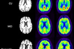
Researchers at the University of Alabama at Birmingham compared early-frame amyloid PET imaging with standard flortaucipir-PET imaging for Alzheimer's disease and found the new method effectively identified brain pathology associated with the disease in three minutes, compared with scans that can take up to three hours.
"Successful development of this approach has the potential to provide information on both amyloid and tau pathology in a single PET session, which may reduce imaging costs and burden on patients and their families," wrote senior author Dr. Jonathan McConathy, PhD, of the University of Alabama at Birmingham.
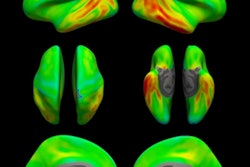
In Alzheimer's disease, abnormal amounts of tau and amyloid proteins accumulate in specific brain regions involved in memory. The PET tracer flortaucipir was designed to detect accumulations of these biomarkers and was approved by the U.S. Food and Drug Administration last year. In standard flortaucipir-PET imaging, the presence of tau and amyloid clumps may be measured between 30 and 120 minutes after injection.
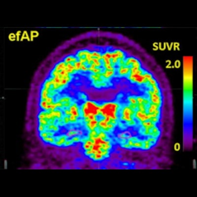
 Comparisons of representative subjects. The first column highlights the hippocampus in pink on volumetric MRI. The second column shows early-frame amyloid PET (efAP) values from dynamic florbetapir-PET. The third column shows standard tau PET. Intensity scales for the PET images are shown as standardized uptake value ratios (SUVRs). Image courtesy of the Journal of Nuclear Medicine.
In this study, the researchers hypothesized that early-frame amyloid PET could detect pathologic tau measurements in amyloid-positive patients on both dynamic amyloid and tau PET scans performed within six months of each other.
They analyzed scans of 63 patients who were amyloid-positive and 57 who were amyloid-negative. The ability of early-frame amyloid PET to stratify positive tau findings was measured using receiver operating characteristics analysis of the area under the curve (AUC). Patients were categorized as amyloid- and tau-positive or negative based on established cutoffs and were predominantly cognitively intact in both groups.
The researchers found that measurements with early frame amyloid PET within the hippocampus within the first three minutes of brain activity postinjection showed the strongest discriminative ability to stratify for tau positivity (AUC, 0.67-0.89 across tau PET regions) in amyloid-positive individuals. In addition, hippocampal early-frame amyloid PET correlated significantly with global tau-PET tauopathy scores in amyloid-positive patients.
"We have shown that [early frame amyloid PET] acquired concurrently with standard amyloid PET study is a strong predictor of tau pathology in amyloid-positive individuals," McConathy and colleagues wrote.
The authors noted the study population included a large proportion of amyloid-positive subjects who were cognitively normal or had mild cognitive impairment and that the optimal brain region for early-frame amyloid PET measurements may be different in individuals with more advanced Alzheimer's disease.
Nonetheless, the successful development of their approach has the potential to provide information on both amyloid and tau pathology in a single PET session, which may reduce imaging costs and burden on patients and their families, the researchers stated.
"Hippocampal [early-frame amyloid PET] can provide additional information to conventional amyloid-PET, including estimation of the likelihood of tau positivity in amyloid-positive individuals," they concluded.