RSNA 2015 is the culmination of a year of progress in advanced visualization, 3D, and computer-aided detection (CAD) technologies that shine a light on tasks that radiologists can now perform better, faster, and often more cheaply. Considering the economic and diagnostic realities that the specialty faces today, it is an urgent task.
The meeting opens with RSNA President Ronald Arenson from the University of California, San Francisco discussing radiology's future and why radiologists must, like the intrepid Star Trek Enterprise crew, go there boldly. Arenson will examine everything from artificial intelligence to sophisticated data mining to dramatic improvements in image quality.
Later in the week, and perhaps closer to earth, Dr. Adam Flanders from Thomas Jefferson University Hospital in Philadelphia will examine an array of available advanced imaging technologies and show what each can offer the radiologist in a hurry.
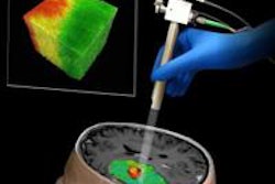
In the head, Italian researchers will discuss how they are using advanced quantitative MRI techniques to map the brain and how various diseases affect it. Meanwhile, authors from Brown University in Providence, RI, will discuss their use of ultrasound CAD to find cases of craniosynostosis (premature fusion of skull bones) that normal prenatal ultrasound can miss -- improving newborns' chances of timely corrective surgery.
In brain cancer, advanced imaging shows that not all gliomas are alike. Investigators applied texture analysis to MRI-based images of glioma multiforme to predict survival in the fast-moving cancer, enabling individualized treatment.
Texture analysis was also tapped to distinguish between two uterine findings that can appear alike: atypical-appearing uterine leiomyomas and leiomyosarcomas, according to a study team from Memorial Sloan-Kettering Cancer Center in New York City.
Bone cancer, too, is meeting its match in advanced visualization. Japanese researchers applied a threshold-based algorithm to SPECT/CT images to detect "hot spots" that represent metastases.
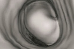
In colorectal cancer screening with CT colonography (CTC), researchers from Massachusetts General Hospital (MGH) in Boston and Harvard University in Cambridge, MA, have delivered two much-needed solutions.
In one study, the MGH team used a CAD scheme on supine-only CTC images without any help from a reader, delivering results that were equivalent to prone and supine imaging without CAD at half the dose. In another study, they developed an electronic cleansing algorithm that removes the colon contents from native CTC images with markedly less artifact than previous attempts.
Japanese researchers also are on the trail in colorectal cancer screening. They developed a gaze-tracking system that monitors where, and for how long, radiologists look for abnormalities on CT colonography images.
In the coronary arteries, Dutch researchers applied an automated coronary calcium scoring technique to an entire Canadian lung cancer screening population, effectively providing two screening tests for the price of one.
Also in cardiac imaging, functional and molecular aortic imaging are remaking care of not just the coronary arteries but the aorta, where evidence of inflammation and potential rupture can be seen years in advance, according to California researchers.
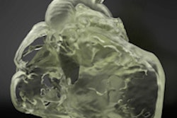
Finally, the burgeoning 3D printing industry, based almost entirely on CT and MR images, is on full display in this year's RSNA sessions.
The fun starts with an overview course by Dr. Frank Rybicki from the University of Ottawa Hospital, who overviews the many courses and scientific sessions devoted to 3D printing applications at the meeting. For the first time, the RSNA has a dedicated space in the Lakeside Center for a 3D printing exhibit, he said.
The volume of cardiovascular imaging applications is large and growing, according to Rybicki, who also leads a course devoted to the subspecialty. Among the beneficiaries are infants with congenital abnormalities and patients with valve disease, to name just two.
Among the dozens of scientific abstracts on 3D printing scheduled for RSNA 2015, one features a group that 3D prints its own customized quality control phantoms.
Another group from Brigham and Women's Hospital in Boston is devoted to improving breast reconstruction via 3D printing, which can address many problems that women face as the result of too many flawed surgeries.
To view RSNA's complete listing of abstracts for this year's scientific and educational program, click here.