Mayo Clinic researchers used iterative reconstruction to acquire half-dose virtual colonoscopy exams and compare them to full-dose exams without scanning patients twice. The technique developed to perform the study gives researchers a more accurate way to compare dose levels between different CT protocols, according to a new study in the Journal of Computer Assisted Tomography.
Accurate CT dose reporting is a perennial issue for radiology researchers trying to evaluate the effectiveness of different low-dose protocols. Because repeating CT scans in the same patient using different scanner settings can rarely be justified, researchers have traditionally resorted to phantom studies to approximate dose differences among different protocols, or divide patients into matched cohorts that undergo different scan protocols to approximate dose differences between groups of patients that are similar but never match up precisely (JCAT, September/October 2012, Vol. 36:5, pp. 560-569).
"Whereas noise reduction has attracted much interest in the radiologic community, there is a dearth of observer performance studies of low-dose CT using noise reduction techniques, largely owing to practical hurdles: Most importantly, full-dose images without noise reduction are needed for comparison purposes to ensure that subtle imaging findings are not obscured or lost," wrote Dr. Joel Fletcher and colleagues from Mayo Clinic in Rochester, MN. "On the other hand, comparison to low-dose images without noise reduction is needed to determine if noise reduction is needed at all."
Simply put, radiation dose and image quality must be compared in every patient and not just between patient groups, because discrepancies in the patient's shape, cardiac output, lesion pathology, and other factors are highly individualized, they wrote. Most important, matched-cohort research studies can't evaluate the impact of noise reduction on reader performance for identifying findings, and even back-to-back full- and half-dose studies cannot control for the effect of phase enhancement on lesion conspicuity.
Also, dose is key in virtual colonoscopy (also known as CT colonography or CTC) studies because screening patients are asymptomatic, "and there is a consequently a greater imperative to minimize the risk of this examination, including radiation dose," Fletcher and colleagues wrote. The goal of the study was to evaluate the dual-source CT (DSCT) technique for obtaining half-dose CT images for use in observer studies evaluating reduced-dose CTC exams.
Phantom arm
To address the dose comparison issue, the researchers used phantoms to acquire images from full-dose dual-source studies in which only data from a single x-ray tube were reconstructed; these images were compared with single-source images acquired at half the dose.
The phantom studies looked at dose and image noise from scans of four acrylic water-filled phantoms ranging from 30- to 45-cm wide. They were scanned using multiple tube potentials with matched dual- and single-source protocols. The results were compared with images from single-tube reconstruction of dual-source data, with those from single-source images acquired at half of the original dose, expressed in CT dose index volume (CTDIvol).
The authors found similar image noise levels for both protocols in all but the largest (≤ 50 cm), confirming that dual-source acquisition with single-tube reconstruction can create half-dose images that can be compared with full-dose images.
Of note, significant differences in image noise were noted in the large 45-cm phantoms for full-dose scans at both 80 and 120 kV. And as expected, image noise increased by about 41% between full- and half-dose images for both single- and dual-source scans, a finding that is in line with current understanding that dose requirements for larger patients are not linear -- and thus these patients require higher dose settings.
Patient arm
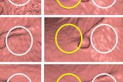
To examine whether half-dose images acquired with iterative reconstruction are equivalent to the diagnostic quality of full-dose images acquired without iterative reconstruction, the researchers examined 30 patients undergoing supine single-source and dose-matched prone dual-source CTC scans, reconstructed using Siemens Healthcare's proprietary sinogram-affirmed iterative reconstruction (SAFIRE) software. Two experienced radiologists scored image quality on both 2D and 3D images.
Images from the patient studies were all reconstructed using a B30 kernel and 1-mm slice thickness at 0.8-mm increments. Across all the image sets, the study team measured image noise in the liver (excluding vessels), subcutaneous fat, and paraspinal muscle for each scan type. The two expert readers subjectively graded image quality, including noise, texture, and diagnostic confidence.
Image noise was similar between half-dose single-source and dual-source images reconstructed from one tube in a dual-source scanner (tube potential 120 kV or higher for phantoms ≤ 40 mm in width (p < 0.05), the study team wrote. Image quality scores were more than 84% concordant between dual- and single-source images. Finally, half-dose prone VC images reconstructed with SAFIRE had at least 84% concordance with standard-dose CTC except for 3D image noise, which yielded only 76% concordance.
Results concordant in phantom and patient arms
"Phantom experiments confirmed that [dual-source] acquisition mode with cross-scatter correction can provide equal dose and image noise compared to the traditional [single-source] scan for small and medium-sized phantoms at tube potentials greater than 100 kV, and that [single-source] images acquired with half dose are equivalent in noise to half-dose [dual-source] images obtained from single-tube reconstructions under similar conditions," Fletcher and colleagues concluded.
Together, the acquisition and reconstruction methods offer a way to create half-dose images with and without noise reduction to compare with full-dose images, enabling lesion conspicuity and observer performance to be evaluated using noise reduction techniques, the study team wrote. For CTC, image quality for evaluating the colon was comparable between full-dose images and half-dose images reconstructed using iterative reconstruction.
|
Study disclosures Some co-authors are employees of Siemens Healthcare, which provided the dual-source scanner via a grant. Both SAFIRE software and the Somatom Flash scanner are Siemens Healthcare technologies. |