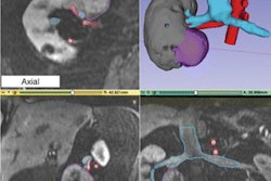
Using a CT scan of a spine, researchers have created a 3D-printed spine replica that could enable improved surgeries for cervical disk implants in patients with degenerative disk disease, according to research published recently in Sensors.
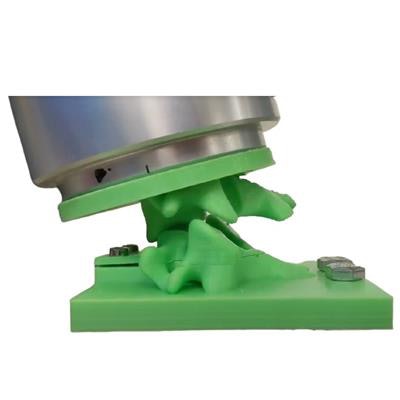
Researchers led by Maohua Lin, PhD, from Florida Atlantic University (FAU), modified this spine replica to include an artificial disk implant and a soft, flexible magnetic sensor array. Artificial intelligence (AI) algorithms were then able to accurately detect both applied loads and postures of the spine after the spine replica was manipulated by a robotic arm.
By using this approach to create a personalized spine "twin," surgeons could preview and compare the potential effects of different surgical interventions prior to surgery.
"Moreover, the novel system could help in determining whether a constrained, semiconstrained, or unconstrained device could be the best fit or even a fusion device," said senior author Dr. Frank Vrionis of Baptist Health's Marcus Neuroscience Institute. "Following surgery, the spine replica could also assist us in estimating whether there is sufficient motion at the operated level and possibly helping us to determine if we need to change the rehabilitation program to prevent calcification and subsequent loss of intended motion."
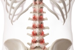
Cervical disk implants are often used to treat degenerative disk disease, but surgeons have to rely primarily on imaging studies -- without any biomechanical data -- to determine if a patient is a candidate for the procedure and to optimize the type of prosthesis, according to the researchers. As a result, patients may experience complications and implant failure.
To help surgeons get a preview for the postoperative effects of an artificial disk implant prior to surgery, the researchers used a patient's CT scan to 3D print a replica of their spine. They then modified the replica to include an artificial disk implant and outfitted it with a soft magnetic sensor array that can detect the location and amplitude of applied loads. The researchers also trained four different machine-learning algorithms to classify the loads, amplitudes, and postures obtained from five different robotically actuated postures.
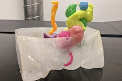
 A 3D-printed replica of a spine could enable surgeons to better plan cervical disk implants. Image courtesy of Florida Atlantic University.
They found that two algorithms -- a random-forest model and an artificial neural network (ANN) -- were able to classify the locations of loads applied 3.25 mm apart with an accuracy of 98.4% and 98.1%, respectively. The ANN also had 94.5% accuracy to classify a location that had received a 10 g load.
After the artificial disk-implant spine replica was subjected to flexion and extension by a robotic arm, the researchers also found that the ANN successfully classified five different postures of the spine with 100% accuracy using the magnetic sensor array.
"All results indicated that the magnetic sensor array has promising potential to generate data prior to invasive surgeries that could be utilized to preoperatively assess the suitability of a particular intervention for specific patients and to potentially assist the postoperative care of people with cervical disk implants," the authors wrote.
In the future, the sensor could potentially also be utilized along with CT exams to address spinal misalignment, according to the researchers.
"Our new approach could provide surgeons with first-hand data to compare the effects of different surgical interventions to treat diseases of the spine before surgery and potentially reduce the rates of complication and failure of artificial disk implantation," said co-author Chi-Tay Tsai, PhD, of FAU in a statement.