PROVIDENCE, RI - A new study from the University of California, Davis shows that real-time molecular imaging is feasible, using a combination of ultrasound technologies and techniques that include transducers with extended bandwidths or multifrequency capabilities, pulse-to-pulse filtering, and radiation force.
The report is among the 929 approved abstracts at the first annual joint conference of the Academy of Molecular Imaging (AMI) and Society of Molecular Imaging (SMI). The four-day forum began Saturday in Providence, RI.
The results indicate that the new technique could provide rapid, sensitive, and selective detection of adherent ultrasound contrast agents in ultrasonic molecular imaging. For the technology, UC Davis is partnering with Siemens Medical Solutions of Malvern, PA, to develop and design transducers that offer both low- and high-frequency arrays in the same device.
A high-frequency array in the center rows of the transducer is surrounded on each side with low-frequency arrays. In one linear multirow array configuration, the center row is comprised of 128 elements operating at 5.24 MHz, while the two outer rows include 64 elements operating at 1.48 MHz.
One other transducer design has low and high frequencies of 2 MHz and 9 MHz, respectively. That configuration also is aligned with 64 elements on the outside of the array and 128 elements on the inside.
Katherine Ferrara, a professor of biomedical engineering at UC Davis, said in her presentation that the design rationale is to "get a high-quality image, to get high/low beams that target the same volume, to deliver a lot of power, and get good thermal performance with minimal software changes in the system."
The result is the ability to transmit and receive from the center rows of the array, or transmit from the outer rows and receive on the inner row. For example, an ultrasound signal of 1.6 MHz is transmitted from the low-frequency arrays and received by the high-frequency array at 7 MHz (T1R7).
The imaging sequence incorporates a T1R7 B-mode image pulse sequence, followed by a 10-second ultrasound radiation force pulse to induce immediate binding, then a second T1R7 B-mode imaging pulse.
"Our approach is to exploit what we consider to be a transient response of a bubble, with the idea that we could transmit two different phases of a pulse -- a negative going first, then a positive pulse going next; or a positive going first and a negative coming after," Ferrara said. "With the negative pulse going first, you get a very rapid collapse of a bubble. That very rapid collapse gives you a very high-pressure signal that has a very broad bandwidth."
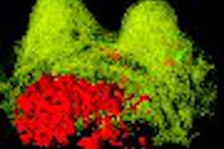
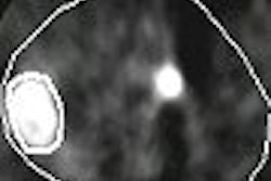
The UC Davis abstract details how the radiofrequency data obtained from the second T1R7 B-mode pulse sequence is averaged and then subtracted from the first T1R7 B-mode sequence. The study found that T1R7 single-cycle pulses suppressed tissue echoes significantly with a bubble contrast-to-tissue ratio (CTR) of 31 dB, compared with a single B-mode pulse CTR of 10 dB.
Without waiting for agent clearance, a targeted bubble contrast-to-free bubble signal ratio of 15-22 dB was obtained from the composite sequence imaging. If a very short pulse is transmitted, a broadband receiver can gather the return signal, and the spatial resolution that is achieved, even after a single pulse, is not necessarily dependent upon the excitation frequency. "You can get away from some of the attenuation effects and improve spatial resolution with that kind of strategy," Ferrara said.
The process also allows for the combination of other techniques that can accomplish real-time molecular imaging. "We transmit a train on pulses, and if we filter or average that train, it allows us to get rid of the precontrast agent," Ferrara said. "If we then apply our radiation force pulse and then another set of transmitted pulses, we can get a similar image ... of targeted agents. We also can subtract those two pieces and end up with an image of just the targeted agent with a signal-to-noise ratio of about 30 dB."
The combination of technique and technology is expected to enhance the capabilities of ultrasound contrast imaging in the near future.
Ferrara noted there are limitations, in that radiation force is less effective in smaller vessels, "so that piece is not as effective and the performance of pulse-to-pulse average will depend on the flow rate."
By Wayne Forrest
AuntMinnie.com staff writer
September 10, 2007
Copyright © 2007 AuntMinnie.com