ANAHEIM, CA - Researchers from the University of Wisconsin-Madison (UWM) developed a whole-body PET protocol with much shorter acquisition times to analyze tumor processes in a new study. But how does it compare to conventional PET? They presented their data at the Society of Nuclear Medicine and Molecular Imaging (SNMMI) annual meeting.
In a comparison of the two PET imaging techniques, whole-body dynamic PET images were rated as "good" or as "excellent" as conventional PET scans in all 19 cases in this study. Whole-body PET had the added advantage of shorter image acquisition times.
"We think whole-body dynamic PET is a promising alternative to conventional [PET] imaging for applications on PET/CT or PET/MRI scanners, which can yield much more robust pharmacokinetic information," said senior author Dr. Steven Cho, associate professor in the nuclear medicine section of UWM's radiology department. "Potentially, we would like to replace single conventional standard-of-care imaging with ... whole-body diagnostic [PET] imaging."
PET's limitations
Standard-of-care PET consists of a single-point composite image usually obtained one hour after injection of FDG. While the modality is relied upon for a number of oncologic applications, it does have its limitations, particularly a lack of robust quantitative data.
Most recently, whole-body dynamic PET imaging has shown to be increasingly feasible in the wake of significant advances in system hardware and technology. The protocol features scans of approximately 30 seconds per bed position, compared with three to five minutes per bed position with the current standard-of-care approach, which is repeated multiple times during an exam.
Especially with the shortened image acquisition times of whole-body dynamic PET, there are tradeoffs, Cho told SNMMI attendees. One obstacle is a greater risk of patient movement between whole-body passes during the exam.
"For each of these passes, because there is a shorter time interval, there is decreased signal-to-noise ratio, so there are noisier images," he noted. "However, [images] potentially can be restored by combining all of the multipass, full-body PET images. The question is: Can we make the whole-body dynamic images similar to standard-of-care images?"
Determining which is best
To answer that question, UWM researchers looked at 13 males and six females (range, 33-68 years) who were scheduled to undergo oncologic standard-of-care PET/CT imaging. Among the 19 subjects, 11 patients received their whole-body dynamic PET scans on a PET/MRI system (GE Signa, GE Healthcare) and eight patients went for a PET/CT (Discovery, GE) exam.
Whole-body dynamic PET imaging began approximately 30 minutes after FDG injection and continued for the 55-minute FDG uptake period. Each set of whole-body dynamic standardized uptake value (SUV) PET images consisted of four to seven whole-body passes.
"Image processing included whole-body dynamic PET images, which were decay-corrected and normalized to create what we call 'SUV images,' which are the averages of all the whole-body dynamic passes," Cho explained. "This enables us to do region-of-interest and time-activity curves, as well as parametric images."
All standard-of-care PET imaging then followed approximately 60 minutes after FDG administration on the PET/CT system.
Two readers who were blinded to which hybrid PET system acquired the images rated the results for image quality based on a five-point scale and motion based on a three-point scale.
'Good or excellent'
Their assessments rated standard PET and whole-body dynamic PET images as either "good" or "excellent" for diagnostic image quality in 18 (95%) of 19 scans. As for motion, 18 scans had "acceptable motion," Cho said. There was one patient who had significant motion during the whole-body dynamic PET image.
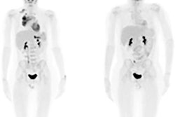
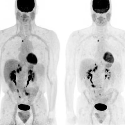
 Maximum intensity projection and transaxial whole-body dynamic (WBD) images and standard-of-care (SOC) images were deemed comparable for cases of head and neck cancer in example 1 and lung cancer in example 2. Images courtesy of Cho et al and SNMMI.
While it was not part of this SNMMI abstract, Cho said the researchers drew regions of interest around normal and tumor structures to look at time-activity curves to see if they could differentiate between malignant and benign processes.
"Whole-body dynamic-averaged SUV images are comparable in image quality to conventional standard-of-care PET images," Cho concluded.