Lung nodule enhancement at CT: multicenter study.
Swensen SJ, Viggiano RW, Midthun DE, Muller NL, Sherrick A, Yamashita K, Naidich
DP, Patz EF, Hartman TE, Muhm JR, Weaver AL
Dept of Diagnostic Radiology, Mayo Clinic and Mayo Foundation, Rochester, MN
55905, USA.
PURPOSE: To test the hypothesis that absence of statistically significant lung
nodule enhancement (< or =15 HU) at computed tomography (CT) is strongly
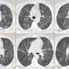
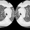
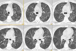
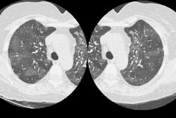
predictive of benignity. MATERIALS AND METHODS: Five hundred fifty lung nodules
were studied. Of these, 356 met all entrance criteria and had a diagnosis. On
nonenhanced, thin-section CT scans, the nodules were solid, 5-40 mm in diameter,
relatively spherical, homogeneous, and without calcification or fat. All
patients were examined with 3-mm-collimation CT before and after intravenous
injection of contrast material. CT scans through the nodule were obtained at 1,
2, 3, and 4 minutes after the onset of injection. Peak net nodule enhancement
and time-attenuation curves were analyzed. Seven centers participated. RESULTS:
The prevalence of malignancy was 48% (171 of 356 nodules). Malignant neoplasms
enhanced (median, 38.1 HU; range, 14.0-165.3 HU) significantly more than
granulomas and benign neoplasms (median, 10.0 HU; range, -20.0 to 96.0 HU; P
< .001). With 15 HU as the threshold, the sensitivity was 98% (167 of 171
malignant nodules), the specificity was 58% (107 of 185 benign nodules), and the
accuracy was 77% (274 of 356 nodules). CONCLUSION: Absence of significant lung
nodule enhancement (< or = 15 HU) at CT is strongly predictive of benignity.
Publication Types:
- Multicenter study
PMID: 10644104, UI: 20106920