J Thorac Imaging 1995;10(4):294-297
CT mosaic pattern of lung attenuation: etiologies and terminology.
Stern EJ, Muller NL, Swensen SJ, Hartman TE
Department of Radiology, Harborview Medical Center, University of Washington, Seattle 98104, USA.
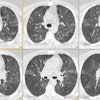
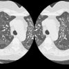
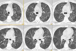
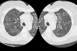
Areas of variable lung attenuation forming a "mosaic pattern" are occasionally seen on computed tomography (CT) or high-resolution CT (HRCT) images of the lungs. This CT mosaic pattern of lung attenuation is a nonspecific finding that can reflect the presence of vascular disease, airway abnormalities, or ground-glass interstitial or air-space infiltrates. However, it is often possible to distinguish among these categories. In small airways disease and pulmonary vascular disease, the pulmonary vessels within the lucent regions of lung are small relative to the vessels in the more opaque lung. In infiltrative diseases, the vessels are more uniform in size throughout the different regions of lung attenuation. The distinction of small airways disease from primary vascular disease requires the use of paired inspiratory/expiratory CT scans. The terms "mosaic perfusion" or "mosaic oligemia" have also been used to describe this heterogeneous pattern of lung attenuation. We believe that the term "mosaic pattern of lung attenuation" is preferable when describing areas of variable lung attenuation because the term "mosaic perfusion" implies pulmonary vascular pathology.
PMID: 8523510, MUID: 96105848