AJR Am J Roentgenol 1995 Oct;165(4):813-816
CT mosaic pattern of lung attenuation: distinguishing different causes.
Stern EJ, Swensen SJ, Hartman TE, Frank MS
Department of Radiology, Harborview Medical Center, University of Washington, Seattle 98104, USA.
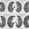
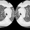
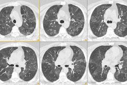
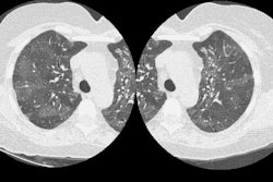
Areas of variable lung attenuation in a lobular or multilobular distribution are occasionally seen on CT or high-resolution CT scans of the lungs [1], although never as a normal finding. This mosaic pattern of lung attenuation presents a challenge to the radiologist when deciding which are the abnormal regions of lung--those of low attenuation, those of high attenuation, or both. We have observed three categories of disease known to cause a CT mosaic pattern of lung attenuation: small-airway disease, vascular lung disease, and infiltrative disease. Diseases from each of these categories can cause similar patterns of mosaic lung attenuation on CT scans. However, it is sometimes possible to distinguish among these categories by using additional CT findings. We illustrate the known causes of a CT mosaic pattern of lung attenuation and highlight distinguishing features.
PMID: 7676972, MUID: 95407448