AJR Am J Roentgenol 1996 Feb;166(2):309-312
Peribronchovascular interstitium of the pulmonary hilum: normal and abnormal findings on thin-section electron-beam CT.
Murata K, Takahashi M, Mori M, Shimoyama K, Nitta N, Mishina A, Matsuo H, Morita R
Department of Radiology, Shiga University of Medical Science, Japan.
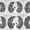
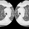
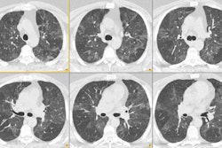
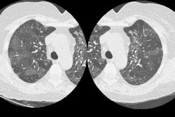
The peribronchovascular interstitium, including bronchial vessels and lymphatic channels, is an important anatomic component of the lung, especially the pulmonary hilum [1-3] (Fig.1). Various pathologic processes involve and spread along this interstitium [4, 5]. Although it has been difficult to accurately evaluate the morphologic changes occurring in the peribronchovascular interstitium with conventional CT or MR imaging [6], spiral CT and electron-beam CT can show these changes clearly and consistently [7, 8]. A judgement as to whether the peribronchovascular interstitium is normal or abnormal is necessary for the CT diagnosis of pulmonary hilar lesions. In this assay, the normal peribronchovascular interstitium and various pathologic processes involving this interstitium are shown as they appear on thin-section electron-beam CT scans.
PMID: 8553936, MUID: 96143098