- Radiology 1998 Jun;207(3):759-65
- Intrapulmonary lesions: percutaneous automated biopsy with
a detachable, 18-gauge, coaxial cutting needle.
Lucidarme O, Howarth N, Finet JF, Grenier PA
Department of Radiology, Hopital de la Pitie-Salpetrere, Paris, France.
PURPOSE: To evaluate a lung biopsy technique in which a detachable, 18-gauge, coaxial guide around a central notched stylet is used as a cutting needle. MATERIALS AND METHODS: The records of 89 consecutive patients (41 women, 48 men; aged 21-86 years) who underwent coaxial percutaneous core biopsy of 91 lung lesions that required needle passage through normal lung tissue (mean lesion size, 33.6 mm; range, 9-80 mm) were studied. Detachable, 18-gauge, coaxial automated cutting needles were used. RESULTS: The mean number of needle passes was 2.5 (range, 1-4). All biopsies yielded sufficient tissue for histopathologic (n = 91) and, if necessary, bacteriologic (n = 12) evaluation (mean core length, 5 mm; range, 1-15 mm). Eighty-nine lesions had definitive diagnoses. Seventy-five lesions were proved to be malignant; seventy (93%) could be accurately diagnosed with coaxial percutaneous core biopsy samples. Fourteen lesions were proved to be benign; 10 (71%) were specifically diagnosed with biopsy samples. Among the 91 biopsies, the overall diagnostic accuracy was 88% (80 of 91 lesions). A pneumothorax occurred in 31 cases (34%), three (3%) of which necessitated placement of a chest tube. Postbiopsy hemoptysis occurred and resolved spontaneously in nine cases (10%). CONCLUSION: This technique provides a core biopsy specimen without the need for an on-site cytopathologist during the procedure. It has a high diagnostic accuracy and an acceptable rate of complications.
Comments:- Comment in: Radiology 1998 Jun;207(3):569-70
- Comment in: Radiology 1999 May;211(2):590-2
PMID: 9609901, UI: 98272821
General > Biopsy
Latest in Pulmonary Imaging
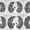
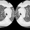
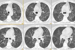
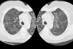
General > ChestCT > Images > Air_Trapping
April 2, 2002

Sponsored