J Thorac Imaging 1995;10(4):227-235
The pathophysiology of airways disease.
Gurney JW
Department of Radiology, University of Nebraska Medical Center, Omaha 68198-1045, USA.
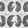
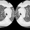
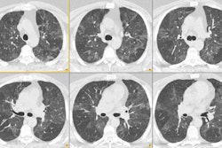
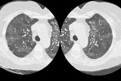
More than 10 million airway branches exist in the normal human lung. Radiographic visualization is < 1% of this total. Many diseases affect the airways, each pathologic insult ultimately resulting in obstruction to airflow. Normally there is little resistance to airflow in the small airways (< 2 mm diameter); thus extensive disease may be present before it becomes clinically evident. Centrilobular emphysema is characterized by dilation and destruction of small airways, whereas bronchiolitis obliterans is characterized by concentric fibrous obliteration of small airways. High-resolution computed tomography, particularly comparison of images at full inspiration and full expiration, is the most sensitive radiographic method with which to image small airways disease.
PMID: 8523504, MUID: 96105842