Bronchogenic Cyst:
View cases of bronchogenic cyst
Clinical:
Bronchogenic cysts account for 50-60% of all mediastinal cysts [2]. A bronchogenic cysts is a bronchopulmonary foregut malformation due to an abnormal budding of the tracheobronchial tree between the 26th and 40th days of gestation which fails to induce surrounding primitive mesenchyme to form normal lung parenchyma. The walls of the cyst are lined by respiratory epithelium, but MUST also contain varying amounts of bronchial cartilage, glands, and smooth muscle to be considered a true bronchogenic cyst. The cyst frequently communicates with the gut or bronchial tree (usually via a fibrous stalk) [3], but if the communication is patent (RARE) this will permit fluid drainage. The lesion may present at any age (although it is usually discovered in adults in the first four decades of life) and the lesion is symptomatic in more than half of the cases [4]. Chest pain is often the most common presenting complaint [3]. Large lesions may produce symptoms of respiratory distress due to compression of adjacent structures or it may become infected. Approximately 70-85% of bronchogenic cysts arise in the mediastinum (usually middle mediastinum in close relationship to the trachea, main bronchi, or in the subcarinal space). Less commonly, the cyst may arise in the posterior mediastinum (about 15% of mediastinal bronchogenic cysts occur in the posterior mediastinum). Up to 15-30% of bronchogenic cysts are intrapulmonary [1,2] - and have a lower lobe predominance. A pulmonary sequestration may be associated with the lesion and is usually distal to the cyst. The lesion can also occur in the diaphragm or pleura [3]. Rarely the bronchogenic cysts may be multiple.
Complications include infection, hemorrhage, and erosion into adjacent structures [5]. Because up to two-thirds of patients may eventually become symptomatic (airway or esophageal obstruction) most lesions are treated surgically to prevent complications and confirm the diagnosis- especially in young patients [2,3]. Lesions can be managed conservatively in asymptomatic adult patients or high risk patients [3]. Exceeding rare is the report of malignancy developing within the cyst wall (rhabdomyosarcoma, pulmonary blastoma, and adenocarcinoma [5].
X-ray:
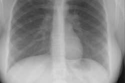
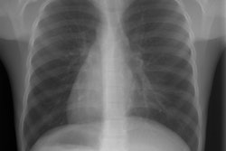
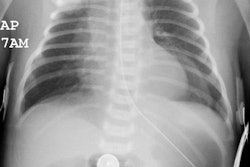
CXR: On plain the cyst appears as a well defined, rounded, fluid filled soft tissue density mass (can see air-fluid level if there is communication to the bronchial tree). Large lesions may displace mediastinal structures or compress bronchi causing atelectasis or distal hyperaeration.
Computed tomography: On CT the cyst is typically a homogeneous water density mass (HU less than 20) with smooth or lobulated sharply marginated borders [3]. Between one-third to 43% of lesions can have homogeneous higher attenuations values if the cyst fluid contains a high protein content, hemorrhage, or calcium and lesion can appear solid. Uncommonly the lesion can have a heterogeneous appearance [3]. Typically the cyst is thin walled and the wall may be below CT resolution. Wall calcification can be seen in about 6% of cases. Milk-of-calcium can be seen in a very small number of cases [3]. Mural enhancement of the cyst wall can be seen following the administration of contrast [3]. No enhancement of the cysts contents should be seen after contrast administration [3].
Intrapulmonary lesions have a similar CT appearance, but are often associated with adjacent areas of low attenuation (due to emphysema) and bandlike opacities (due to fibrotic change) [4].
MRI: On MRI the cyst typically has a homogeneous high signal intensity that is equal to or greater than CSF on T2 weighted images. T1 characteristics vary with the protein content, with either homogeneous low (low protein) or high (high protein) signal. A fluid-fluid layer within the lesion confirms its cystic nature.
REFERENCES:
(1) AJR 1997; Congenital thoracic lesions that mimic neoplastic disease on chest radiographs of adults. 168(3), 763-769 (No abstract available)
(2) Chest 1997; Strollo DC, et al. Primary mediastinal tumors. Part II: Tumors of the middle and posterior mediastinum. 112:1344-1357
(3) Radiology 2000; McAdams HP, et al. Bronchogeic cyst: Imaging features with clinical and histopathologic correlation. 217: 441-446
(4) AJR 2002; Yoon YC, et al. Intrapulmonary bronchogenic cyst: CT and pathologic findings in five adult patients. 179: 167-170
(5) Radiol Clin N Am 2005; Paterson A. Imaging evaluation of congenital lung abnormalities in infants and children. 43: 303-323