Marfan's Syndrome
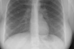
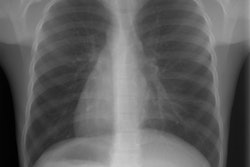
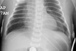
View cases of Marfan's syndrome
Clinical:
Marfan's syndrome is a connective tissue disorder that affects both seexes equally [3]. It is inhereted as an autosomal dominant disorder (70-85% of cases) with high penetrance, but expression is highly variable. Approximately 15% of cases are felt to be due to spontaneous mutation [1]. The disorder has been linked to a fibrillin gene defect on chromosome 15 [2,3]. Marfan's primarily affects the skeletal, cardiovascular, and occular systems. Skeletal abnormalities are almost always present and include arachnodactyly, long limbs, pectus excavatum (or carinatum), joint laxity, and scoliosis. Cardiovascular abnormalities are found in 60-90% of patients. Cystic medial necrosis associated with the syndrome results in weakening of the aortic wall typically resulting in ascending aortic aneurysms. Annuloaortic ectasia occurs in 60-80% of adults with Marfans syndrome and refers to uniform dilatation of all three sinuses of Valsalva, with extension into the ascending aorta and obliteration of the normal sinotubular ridge [3]. Associated valvular dysfunction is also common secondary to dilatation of the aortic root [1]. Occular abnormalities are seen in 50-80% of patients- most commonly subluxation of the lens (ectopia lentis). Pulmonary abnormalities are not a major feature of Marfan syndrome and are found in about 10% of patients [1]. Spontaneous pneumothorax occurs in 4 to 11% of patients (which is several hundred times the incidence in the general population) and is usually the result of apical bleb/bullous disease [1]. Patchy emphysematous changes and bronchiectasis have also been described in patients with Marfan's [1].Regardless of the patient's symptoms, elective replacement of the aortic root is indicated before critical enlargement or dissection occurs - surgery has been suggested for an aortic root diameter of 45mm or if there is a growth rate greater than 0.5 cm/year [3].
X-ray:
On MR imaging of aortic aneurysms associated with Marfan syndrome, there is dilatation of the aortic root associated with complete effacement of the sinotubular junction (remember, this is not seen in atherosclerotic aneurysms until late). When viewed in an oblique coronal section this produces an "onion bulb" or "pear-shaped" appearance to the aortic root and this is considered to be characteristic of the disorders. Cine gradient images can be used to demonstrate regurgitant flow associated with valvular dysfunction.REFERENCES:
(1) J Thorac Imaging 1992; Tanoue LT. Pulmonary involvement in collagen vascular disease: A review of the pulmonary manifestations of the Marfan syndrome, ankylosing spondylitis, Sjogren's syndrome, and relapsing polychondritis. 7 (2): 62-77
(2) Radiology 1997; Kawamoto S, et al. Thoracoabdominal aorta in Marfan syndrome: MR imaging findings of progression of vasculopathy after surgical repair. 203(3): 727-732
(3) Radiographics 2010; Kimura-Hayama ET, et al. Uncommon congenital and acquired aortic diseases: role of multidetector CT angiography. 30: 79-98


