Monday, November 28 | 9:30 a.m.-10:30 a.m. | M3-SSMK04-3 | Room N228
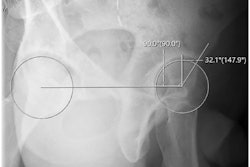
In this musculoskeletal imaging session, researchers will discuss the use of a commercially available artificial intelligence (AI) algorithm for identifying anatomical landmarks of hip dysplasia on anteroposterior pelvis x-rays.In addition to providing extensive time savings, integration of the algorithm could help in places without access to board-certified radiologists or orthopedic surgeons to conduct the measurements, the researchers suggest.
A group led by orthopedic surgeons at the University of Texas Southwestern Medical Center in Dallas tested an algorithm (IB Lab HIPPO, Image Biopsy Lab) in 130 patients with hip dysplasia compared with measurements by three trained readers. The algorithm performs six measurements associated with hip dysplasia.
Among 256 hips with AI outputs, all six hip AI measurements were successfully obtained, according to the study. The AI-reader correlations were generally fair to excellent, with the most widely used measurements for hip dysplasia diagnosis (lateral center edge angle and Tönnis angle) demonstrating good to excellent intermethod reliability.
In addition, the median reading time for the three readers and the AI algorithm were 212, 131, 734, and 41 seconds, respectively. On average, for a given patient, the AI algorithm performed reads that were 80.4%, 70.1%, and 94.4% faster than reader 1, reader 2, and reader 3, according to the findings.
“This study validated that AI-based trained software demonstrated significant time savings in reliable radiographic assessment of patients with hip dysplasia,” noted UT Southwestern resident Holden Archer, who will present the study.