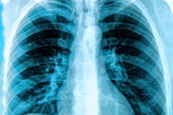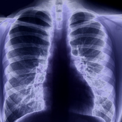
An artificial intelligence (AI) algorithm can accurately identify pneumothorax on chest x-rays by comparing new cases with a database of patients who have similar images and known findings, according to research being presented this week at the Vector Institute's Evolution of Deep Learning Symposium in Toronto.
A team of researchers from the University of Waterloo in Canada developed an AI model that analyzes chest x-rays of new patients by comparing the images with a database of over 500,000 chest x-rays with known diagnoses, including 30,000 exams with pneumothorax. The algorithm was able to identify 75% of the pneumothorax cases, an improvement over an average of less than 50% traditionally diagnosed on chest x-rays, according to the researchers.
In a project funded by nonprofit company Vector Institute, the University of Waterloo researchers are working with Toronto healthcare organization University Health Network (UHN) to increase the accuracy of the software to 90%. The goal by next year is to then integrate the algorithm into Coral Review, a software system developed and used at UHN-affiliated hospitals.
If successful at UHN, the model would then be offered to other hospitals using Coral Review, according to the researchers. They believe the system could offer radiologists a "computational second opinion" in Coral, or it could be used to prioritize x-rays to be looked at first.
In the future, the researchers plan to apply the core algorithm to search for other conditions such as pneumonia and chronic obstructive pulmonary disease.