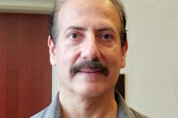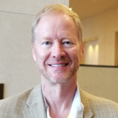
AUSTIN, TX - Dr. Bradley Erickson, PhD, of the Mayo Clinic in Rochester, MN, discussed several important trends in artificial intelligence (AI) for medical imaging in a video interview at the Society for Imaging Informatics in Medicine's Conference on Machine Intelligence in Medical Imaging (C-MIMI).
Moving beyond the early hype that AI could potentially outperform radiologists, C-MIMI 2019 featured an increasing emphasis on the practical implications of applying AI in radiology, said Erickson, who served as co-chair for the conference.
"There's been a lot more focus in this meeting on 'How do we integrate the results [of AI] with the practice of radiology?' " he said. "So it's not by any means about replacing the radiologist. It's 'What does [AI] mean for the workflow of the radiologist, how does it improve the quality of what we can do, [and] how does it improve the patient care experience?' "
Other key trends include the use of AI in nonradiology applications such as in cardiology, ophthalmology, and dermatology, according to Erickson. Importantly, several presentations highlighted efforts to facilitate training of algorithms using data from multiple sites but without requiring the actual patient data to be shared among institutions.
"I think there's a lot of interest in how to do that effectively just because of the concerns about patient privacy and the need for good generalization across multiple institutions," he said.
Erickson also shared his thoughts on a number of other AI topics, including triage applications such as for reducing the mammography workload for radiologists, as well as how AI tools can demonstrate value in radiology.
Dr. Bradley Erickson, PhD, of the Mayo Clinic and co-chair of C-MIMI 2019.


![A normal mammogram confirmed by three-year radiologic follow-up illustrates reader-marked regions of interest (ROIs) during (A) unaided (round 1) and (B) artificial intelligence (AI)–assisted (round 2) reading. Each colored dot represents an ROI for recall by a human reader. Readers could mark more than one ROI per case, represented by multiple dots of the same color. During AI-assisted reading, the AI system displayed three visible prompts: two with suspicion of malignancy scores of 35% (left mediolateral oblique [L MLO] and craniocaudal [L CC]) and one with a suspicion of malignancy score of 10% (right craniocaudal [R CC]), shown as polygonal overlays. Without AI, six of 10 readers (60%) marked a false-positive ROI. With AI assistance, this fell to two of 10 (20%). R MLO = right mediolateral oblique.](https://img.auntminnie.com/mindful/smg/workspaces/default/uploads/2026/07/2026-07-14-radiology-mammogram-ai-auto-bias.H0bYO8QlWs.jpg?auto=format%2Ccompress&fit=crop&h=100&q=70&w=100)


![A normal mammogram confirmed by three-year radiologic follow-up illustrates reader-marked regions of interest (ROIs) during (A) unaided (round 1) and (B) artificial intelligence (AI)–assisted (round 2) reading. Each colored dot represents an ROI for recall by a human reader. Readers could mark more than one ROI per case, represented by multiple dots of the same color. During AI-assisted reading, the AI system displayed three visible prompts: two with suspicion of malignancy scores of 35% (left mediolateral oblique [L MLO] and craniocaudal [L CC]) and one with a suspicion of malignancy score of 10% (right craniocaudal [R CC]), shown as polygonal overlays. Without AI, six of 10 readers (60%) marked a false-positive ROI. With AI assistance, this fell to two of 10 (20%). R MLO = right mediolateral oblique.](https://img.auntminnie.com/mindful/smg/workspaces/default/uploads/2026/07/2026-07-14-radiology-mammogram-ai-auto-bias.H0bYO8QlWs.jpg?auto=format%2Ccompress&fit=crop&h=112&q=70&w=112)