At the upcoming American Brachytherapy Society (ABS) meeting in Orlando, FL, computer-aided detection (CAD) and radiation therapy firm iCAD, plans to highlight first-year data from a clinical trial that is evaluating the company's Xoft system for treating early-stage breast cancer.
Dr. A.M. Nisar Syed of Long Beach Memorial Medical Center in Long Beach, CA, and colleagues are comparing traditional external-beam radiation therapy (EBRT) with intraoperative radiation therapy (IORT) using the Xoft system. Syed's group has found early results from the study show that IORT delivers good to excellent cosmetic results in 94% of cases and very few low-grade adverse events in select patients with early-stage breast cancer, according to iCAD.



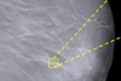


![A normal mammogram confirmed by three-year radiologic follow-up illustrates reader-marked regions of interest (ROIs) during (A) unaided (round 1) and (B) artificial intelligence (AI)–assisted (round 2) reading. Each colored dot represents an ROI for recall by a human reader. Readers could mark more than one ROI per case, represented by multiple dots of the same color. During AI-assisted reading, the AI system displayed three visible prompts: two with suspicion of malignancy scores of 35% (left mediolateral oblique [L MLO] and craniocaudal [L CC]) and one with a suspicion of malignancy score of 10% (right craniocaudal [R CC]), shown as polygonal overlays. Without AI, six of 10 readers (60%) marked a false-positive ROI. With AI assistance, this fell to two of 10 (20%). R MLO = right mediolateral oblique.](https://img.auntminnie.com/mindful/smg/workspaces/default/uploads/2026/07/2026-07-14-radiology-mammogram-ai-auto-bias.H0bYO8QlWs.jpg?auto=format%2Ccompress&fit=crop&h=112&q=70&w=112)


