Dear Advanced Visualization Insider,
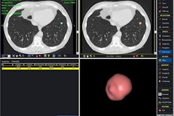
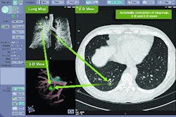
Lung computer-aided detection (CAD) software shows promise as a second reader of low-dose thoracic CT studies. However, the software can also produce a large number of false-positive findings, as well as some troubling false negatives, according to researchers from the University Health Network in Toronto.
The good news is that CAD was able to detect nodules that had been overlooked by the radiologist. The group's presentation at the Computer-Assisted Radiology and Surgery meeting in Berlin, covered by staff writer Eric Barnes, is the subject of our Insider Exclusive article this month.
As an Advanced Visualization Insider subscriber, you have access to this article before it is published for the rest of our AuntMinnie.com members. To read more details about the Canadian lung CAD research, click here.
Is there a topic you'd like to see covered, or are you interested in submitting an article to AuntMinnie.com? Please feel free to drop me a line.