Dear AuntMinnie Member,
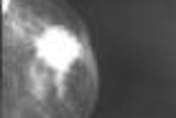
As computer-aided detection (CAD) software begins to permeate the clinical mainstream, it is demonstrating its value in a number of key applications. Some breast imaging specialists, in fact, wouldn't dream of reading mammograms without it.
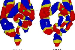
CAD's developing role in medical imaging's armamentarium is the focus of a new two-part series in our Advanced Visualization Digital Community. Written by contributing writer Wayne Forrest, the series explores the growing use of CAD technology, from breast imaging to virtual colonoscopy.
Part I of the series discusses how CAD is making it easier for radiologists to review breast MR images. Breast MRI CAD not only improves accuracy but also streamlines workflow and helps practices standardize their reports, according to those who are using it.
The reimbursement situation for CAD is also improving, with the technology in recent months receiving a thumbs-up from a major payor that stated that CAD is now "a medically necessary adjunct to mammography."
You'll find the rest of the story by clicking here. And be sure to come back tomorrow for part II, which will cover reimbursement, workflow, and CAD's growing role in multidetector-row CT and virtual colonoscopy -- all in your Advanced Visualization Digital Community, at av.auntminnie.com.