Dear Advanced Visualization Insider,
3D printing has become so commonplace that some surgeons won't do a complex procedure without a 3D-printed model in hand. But the technology is really just one way to look at a 3D volume, according to a talk at the recent 3DHeals meeting in San Francisco. The future of 3D -- and in some cases the present -- will see clinicians shifting seamlessly from raw image data to holography to a 3D-printed model when needed, investigators said.
To engage with the data on all of those levels, however, you'll need higher resolution than what's contained in the stereolithography (STL) file format that's most often used for 3D printing. And you'll need better ways to explore the data. Check out one firm's solution in our Insider Exclusive.
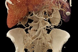
In computer-aided detection (CAD), a generation of CAD algorithms has culminated in impressive results in several anatomic regions -- such as the lungs and the colon, to name two examples where CAD routinely outperforms human readers. But what if these applications operated more like the sick bay in "Star Trek," where Dr. McCoy could use noninvasive multiorgan analysis to quickly determine the patient's condition within minutes after a brutal attack by aliens?
To be sure, the engineers and radiologists at the U.S. National Institutes of Health aren't there yet, but they're trying. In a presentation at last week's International Society for Computed Tomography (ISCT) meeting, NIH senior investigator Dr. Ronald Summers, PhD, discussed the agency's CAD projects aimed at analyzing CT scans of the abdomen, looking for everything from osteoporosis to small-bowel tumors to polyps in one fell swoop. They haven't put together the one-button one-stop pan-CAD application to launch automated abdomen analysis, but they're well on their way.
Then there's the buzz around cinematic rendering, the photorealistic technique that ups the ante on advanced data rendering to provide lifelike images that avoid looking like computer-generated images. The resulting images are almost indistinguishable from real anatomy, judging by the reaction at last week's ISCT meeting. Find out how radiologists from Johns Hopkins University do it by clicking here.
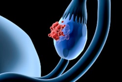
Of course, 3D printing does more than create educational and training models. It can build organs, and as the design, printing technology, and materials to do it evolve, we can expect to see a lot more bioprinted organs. For example, investigators at Northwestern University have succeeded in printing mouse ovaries that house viable eggs that yield healthy offspring, according to a new report. Get the rest of the story here.
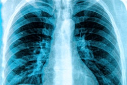
Meanwhile, researchers from Japan are advancing the capabilities of the standard chest x-ray with an algorithm that tells doctors whether an exposure was positioned correctly. They hope to use the technology to flag and correct image orientation errors that occur with wireless digital radiography (DR) as well as with larger DR panels.
Finally, a project from Southern Illinois University takes radiology residents and throws them headlong into the frenetic, fast-paced world of a busy reading room via virtual reality simulation. Complete immersion simulation gives teachers a new tool they can use to train and evaluate many aspects of being a radiologist, presenters said at the recent American College of Radiology (ACR) annual meeting. What are you waiting for? Jump in here.
But wait, there's more. We invite you to scroll through the links below for the rest of the news in advanced visualization, 3D printing, and computer-aided detection, all here in your Advanced Visualization Community.