Medical imaging's greenhouse gas emissions are primarily determined by the carbon intensity of state power grids rather than scanner usage alone, with grid decarbonization accounting for 76% of state variation in emissions. Healthcare facilities should prioritize grid decarbonization and energy procurement strategies alongside traditional imaging stewardship to reduce their environmental footprint.
- Grid carbon intensity accounts for 76.3% of state variation in per-beneficiary greenhouse gas emissions from MRI and CT imaging
- MRI uses significantly more electricity than CT, drawing about 20 kWh per exam compared to 1.2 kWh for CT, though CT's higher volume creates a large overall footprint
- Emissions vary dramatically by state, ranging from 0.05 MT CO₂e in Vermont to 4.82 MT CO₂e in Missouri based on local power grid composition
- States with high carbon intensity should prioritize grid decarbonization and renewable energy procurement as complementary strategies to reducing imaging utilization
U.S. imaging leaders may need to look to their respective state’s power grid for energy consumption by CT and MRI scanners, according to research published August 26 in the American Journal of Roentgenology.
Variation in grid carbon intensity by state drives per-beneficiary greenhouse gas emissions, wrote researchers led by Robert French MD, from the Mayo Clinic in Phoenix.
“The findings highlight the role of grid decarbonization and energy procurement strategies in complementing imaging stewardship for reducing imaging's environmental footprint,” French and colleagues wrote.
In medical imaging, MRI and CT are eyed for their energy consumption. A single MRI examination draws about 20 kWh of electricity, while CT draws about 1.2 kWh of electricity per exam. While CT uses less electricity, its “far-greater” annual volumes lead to the modality’s large energy footprint in radiology.
Choropleth map of state-level imaging-associated greenhouse gas emissions per 1,000 Medicare Part B fee-for-service beneficiaries in 2022. Darker shades indicate higher per-beneficiary greenhouse gas emissions. District of Columbia (not shown) is in 3-4 MTCO2e per 1,000 range. MT CO₂e = metric tons of carbon dioxide equivalentARRS
The researchers noted that MRI- and CT-associated greenhouse gas emissions are also decided by the carbon intensity of the power grid supplying electricity. This, in turn, reflects local fuel mixtures, they wrote.
French and co-authors studied the relative contributions of imaging use and grid carbon intensity to state variation in per-beneficiary greenhouse gas emissions tied to MRI and CT. The study included data collected in 2022 from federal datasets for Medicare Part B fee-for-service beneficiaries.
While CT was used more than MRI in 2022, MRI had higher associated estimated electricity use and greenhouse gas emissions.
Energy use from CT, MRI for calendar year 2022 | ||
Measure | CT | MRI |
National use per 1,000 beneficiaries | 805 exams | 271 exams |
Associated estimated electricity use | 28,236 MWh | 157,734 MWh |
Associated estimated greenhouse gas emissions | 9,991 MT CO₂e | 54,052 MT CO₂e |
The team also reported a national estimated 2.19 MT CO₂e for greenhouse gas emissions per 1,000 beneficiaries. This ranged from 0.05 MT CO₂e in Vermont to 4.82 MT CO₂e in Missouri.
Finally, the researchers found that carbon intensity accounted for 76.3% of state variation in per-beneficiary greenhouse gas emissions, utilization-weighted electricity use for 16.3%, and their covariance for 7.4%.
The study authors suggested that states with high carbon intensity could prioritize decarbonization and other energy gathering efforts. However, they noted that this is complementary to reducing low-value imaging and improving energy efficiency for MRI and CT scanners.
“Indeed, states with both high imaging utilization and high grid carbon intensity could serve as particular targets of sustainability efforts,” the authors wrote.
Read the full study here.
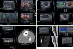
![Examples of ultrasound findings and techniques. (A) Images in a 39-year-old male patient with a mass in the left thigh. The mass is heterogeneous on the B-mode US image (compared with the patient in D) and showed increased microvascularity (superb microvascular imaging [SMI]) and shear-wave elastography (SWE) values. Undifferentiated pleomorphic sarcoma was diagnosed at biopsy (with pleomorphic rhabdomyosarcoma in surgical specimen). (B) Images in an 18-year-old male patient with a mass in the left leg. The mass is hypoechoic on the B-mode image, with no other findings suggestive of malignancy. The lesion is in contact with the cortex of the tibia, which is slightly irregular. CT revealed a doubtful anteromedial tibial erosion. The microvascular study demonstrated high vascularization, suggestive of malignancy. Periosteal Ewing sarcoma was diagnosed with both histologic and immunohistochemical confirmation. (C) Images in a 69-year-old female patient with a lump growing on the outside of the left leg. Multiple SWE examinations were performed (please note the high values obtained in the measurements, whereas the color map highlights the stiffness relative to adjacent tissues). SMI showed areas of increased vascularization to target for sampling. Undifferentiated spindle cell sarcoma was diagnosed at biopsy, with residual leiomyosarcoma in the surgical specimen after neoadjuvant therapy. (D) Images in a 56-year-old female patient with a mass in the right thigh. The mass is heterogeneous at both B-mode ultrasound (similar to patient A) and MRI (coronal T2-weighted spectral attenuated inversion recovery [SPAIR]; T1-weighted pre-contrast and postcontrast imaging), which even shows uptake after the administration of paramagnetic contrast material, which is traditionally suggestive of malignancy. Low values at SMI and elastography are suggestive of benignity. Spindle cell lipoma was diagnosed at biopsy, with atypical spindle cell lipomatous tumor in the surgical specimen.](https://img.auntminnie.com/mindful/smg/workspaces/default/uploads/2026/08/images-radiol250278fig2.APCFLSvX6p.jpg?auto=format%2Ccompress&fit=crop&h=100&q=70&w=100)


![Examples of ultrasound findings and techniques. (A) Images in a 39-year-old male patient with a mass in the left thigh. The mass is heterogeneous on the B-mode US image (compared with the patient in D) and showed increased microvascularity (superb microvascular imaging [SMI]) and shear-wave elastography (SWE) values. Undifferentiated pleomorphic sarcoma was diagnosed at biopsy (with pleomorphic rhabdomyosarcoma in surgical specimen). (B) Images in an 18-year-old male patient with a mass in the left leg. The mass is hypoechoic on the B-mode image, with no other findings suggestive of malignancy. The lesion is in contact with the cortex of the tibia, which is slightly irregular. CT revealed a doubtful anteromedial tibial erosion. The microvascular study demonstrated high vascularization, suggestive of malignancy. Periosteal Ewing sarcoma was diagnosed with both histologic and immunohistochemical confirmation. (C) Images in a 69-year-old female patient with a lump growing on the outside of the left leg. Multiple SWE examinations were performed (please note the high values obtained in the measurements, whereas the color map highlights the stiffness relative to adjacent tissues). SMI showed areas of increased vascularization to target for sampling. Undifferentiated spindle cell sarcoma was diagnosed at biopsy, with residual leiomyosarcoma in the surgical specimen after neoadjuvant therapy. (D) Images in a 56-year-old female patient with a mass in the right thigh. The mass is heterogeneous at both B-mode ultrasound (similar to patient A) and MRI (coronal T2-weighted spectral attenuated inversion recovery [SPAIR]; T1-weighted pre-contrast and postcontrast imaging), which even shows uptake after the administration of paramagnetic contrast material, which is traditionally suggestive of malignancy. Low values at SMI and elastography are suggestive of benignity. Spindle cell lipoma was diagnosed at biopsy, with atypical spindle cell lipomatous tumor in the surgical specimen.](https://img.auntminnie.com/mindful/smg/workspaces/default/uploads/2026/08/images-radiol250278fig2.APCFLSvX6p.jpg?auto=format%2Ccompress&fit=crop&h=167&q=70&w=250)

![Examples of ultrasound findings and techniques. (A) Images in a 39-year-old male patient with a mass in the left thigh. The mass is heterogeneous on the B-mode US image (compared with the patient in D) and showed increased microvascularity (superb microvascular imaging [SMI]) and shear-wave elastography (SWE) values. Undifferentiated pleomorphic sarcoma was diagnosed at biopsy (with pleomorphic rhabdomyosarcoma in surgical specimen). (B) Images in an 18-year-old male patient with a mass in the left leg. The mass is hypoechoic on the B-mode image, with no other findings suggestive of malignancy. The lesion is in contact with the cortex of the tibia, which is slightly irregular. CT revealed a doubtful anteromedial tibial erosion. The microvascular study demonstrated high vascularization, suggestive of malignancy. Periosteal Ewing sarcoma was diagnosed with both histologic and immunohistochemical confirmation. (C) Images in a 69-year-old female patient with a lump growing on the outside of the left leg. Multiple SWE examinations were performed (please note the high values obtained in the measurements, whereas the color map highlights the stiffness relative to adjacent tissues). SMI showed areas of increased vascularization to target for sampling. Undifferentiated spindle cell sarcoma was diagnosed at biopsy, with residual leiomyosarcoma in the surgical specimen after neoadjuvant therapy. (D) Images in a 56-year-old female patient with a mass in the right thigh. The mass is heterogeneous at both B-mode ultrasound (similar to patient A) and MRI (coronal T2-weighted spectral attenuated inversion recovery [SPAIR]; T1-weighted pre-contrast and postcontrast imaging), which even shows uptake after the administration of paramagnetic contrast material, which is traditionally suggestive of malignancy. Low values at SMI and elastography are suggestive of benignity. Spindle cell lipoma was diagnosed at biopsy, with atypical spindle cell lipomatous tumor in the surgical specimen.](https://img.auntminnie.com/mindful/smg/workspaces/default/uploads/2026/08/images-radiol250278fig2.APCFLSvX6p.jpg?auto=format%2Ccompress&fit=crop&h=112&q=70&w=112)