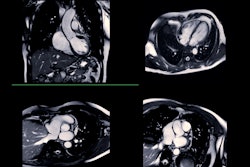
AuntMinnie
  • Europe
  • Clinical News
  • Informatics
  • Industry News
  • Practice Management
  • Education
  • Subspecialties
  • More
  • Sign In
  • Board Review
  • CME
  • Careers
  • Cases
  • Conferences
  • Videos
  • Webinars
  • Podcasts
  • Advertising
  • Buyer's Guide
  • Vendors
  • Minnies
Clinical News
  • CT
  • Digital X-Ray
  • Interventional
  • Molecular Imaging
  • MRI
  • Radiation Oncology/Therapy
  • Ultrasound
  • Womens Imaging
Imaging Informatics
  • Advanced Visualization
  • Enterprise Imaging
  • AI
  • Cybersecurity
Industry News
  • Installations
  • Product News
  • Regulatory
  • M&A
  • Market Analysis
Practice Management
  • Administration
  • Associations
  • Careers
  • Equity & Inclusion
  • Legislation
  • Medicolegal
  • Patient Safety
  • Radiologic Technologist
  • Service
  • Salary Scan
Resources
  • Board Review
  • CME
  • Careers
  • Cases
  • Conferences
  • Videos
  • Webinars
  • Podcasts
  • Advertising
  • Buyer's Guide
  • Vendors
  • Minnies
User Tools
  • Sign In
More
  • About Us
  • Advertising
  • Help
Follow AuntMinnie
Facebook iconTwitter X icon LinkedIn icon
  • Europe
  • Clinical News
    • CT
    • Digital X-Ray
    • Interventional
    • Molecular Imaging
    • MRI
    • Radiation Oncology/Therapy
    • Ultrasound
    • Womens Imaging
  • Informatics
    • Advanced Visualization
    • Enterprise Imaging
    • AI
    • Cybersecurity
  • Industry News
    • Installations
    • Product News
    • Regulatory
    • M&A
    • Market Analysis
  • Practice Management
    • Administration
    • Associations
    • Careers
    • Equity & Inclusion
    • Legislation
    • Medicolegal
    • Patient Safety
    • Radiologic Technologist
    • Service
    • Salary Scan
  • Education
    • Medical Students
    • Residents/Fellows
    • Scott Williams
  • Subspecialties
    • Breast Imaging
    • CV
    • Chest
    • Emergency
    • GI
    • GU
    • Head & Neck
    • Interventional
    • Physics
    • MSK
    • Neuro
    • Nuclear
    • Pediatric
    • Radiation Oncology
  • More
  • Board Review
  • CME
  • Careers
  • Cases
  • Conferences
  • Videos
  • Webinars
  • Podcasts
  • Advertising
  • Buyer's Guide
  • Vendors
  • Minnies
  • Sign In
Follow AuntMinnie
Facebook iconTwitter X icon LinkedIn icon

Abraham Kim

[email protected]
MRI
VR, AR facilitate planning for brain cancer surgery
Tuesday, November 27 | 10:50 a.m.-11:00 a.m. | SSG10-03 | Room E353AClinicians can use virtual reality (VR) and augmented reality (AR) technology to view 3D models of MRI scans during presurgical planning for brain tumor resection, according to this Tuesday session.
November 5, 2018
MRI
3D MRA enables presurgical survey of prostate arteries
Monday, November 26 | 11:50 a.m.-12:00 p.m. | SSC15-09 | Room E352Researchers in this presentation will examine the viability of using 3D contrast-enhanced MR angiography (MRA) to visualize prostate arteries before performing an interventional radiology procedure.
November 5, 2018
CT
Cinematic rendering bolsters visualization of acute trauma
Monday, November 26 | 11:20 a.m.-11:30 a.m. | SSC04-06 | Room S504ABIn this Monday presentation, researchers from Canada will detail their initial experience with cinematically rendered images in the context of acute trauma.
November 5, 2018
2017 11 07 18 37 9362 Roadies Ribbon 400
CT
3D depth camera fine-tunes patient positioning on CT
Monday, November 26 | 10:50 a.m.-11:00 a.m. | SSC13-03 | Room N230BAutomated patient positioning for CT exams using a 3D camera may be more accurate than conventional positioning by radiologic technologists, according to researchers from Germany and Switzerland.
November 5, 2018
Advanced Visualization
How to apply virtual and augmented reality in radiology
Sunday, November 25 | 12:30 p.m.-1:00 p.m. | IN007-EC-SUA | Lakeside, IN CommunityParticipants in this hands-on, fully immersive educational presentation will have the opportunity to learn about a wide range of applications for virtual and augmented reality technology in radiology.
November 5, 2018
MRI
Is 3D MRI or 3D CT best for assessing hip joints?
Sunday, November 25 | 10:45 a.m.-10:55 a.m. | SSA15-01 | Room E353B3D virtual models based on pelvic MRI scans can facilitate presurgical planning for hip joint surgery about as well as 3D CT models can, according to researchers from Switzerland.
November 5, 2018
Advanced Visualization
3D printing enhances training for cleft lip repair
A 3D-printed skull base provided surgical residents with the opportunity to simulate cleft lip repair on a physical model -- a more interactive alternative to the standard practice of training in virtual reality, according to an article published online November 1 in JAMA Facial Plastic Surgery.
November 1, 2018
2018 03 14 00 19 5162 3 D Printing 400
MRI
Heart valve-tracking algorithm boosts viability of 4D MRI
An automated heart valve-tracking algorithm halved the amount of time and variability involved in blood-flow quantification with 4D MRI, according to an article published online October 30 in Radiology. The improved efficiency may help convince clinicians that cardiac 4D MRI is useful for assessing valvular heart disease.
November 1, 2018
2018 08 27 23 46 8725 Heart Ekg 400
CT
Road to RSNA 2018: CT Preview
The scientific presentations focusing on CT at this year's RSNA conference speak to the modality's impressive resilience and continued development. Artificial intelligence (AI) algorithms, computer-aided detection (CAD), and updated protocols that are helping to reduce radiation dose, as well as the results from early studies of ultrahigh-resolution CT scanners, will be key topics discussed at the meeting.
October 31, 2018
2017 11 02 17 42 1107 Road To Rsna 400
Image-Guided Surgery
Mobile CT primed for image-guided brachytherapy
Thursday, November 29 | 11:50 a.m.-12:00 p.m. | SSQ17-09 | Room N229Mobile CT scanning combined with an artifact-reduction algorithm may enhance the visualization of soft tissue for radiotherapy, according to this Thursday session.
October 31, 2018
Previous Page
Page 36 of 63
Next Page
Top Stories
2025 07 18 Silicosis Thumbnail
Molecular Imaging
PET/CT scans show lung damage in stone workers
The metabolic activities occurring in the lungs of these patients have been unknown.
Ctca
CT
SCCT: Study explores diagnostic accuracy of CCTA
Atherosclerosis Clot
CT
SCCT: AI predicts coronary artery calcium scores from chest x-rays
Heart Valves 3 D Base Thumbnail
CT
SCCT: CCTA model shows plaque volume changes in chest pain patients
When CT meets the power of AI everyone benefits.
Sponsor Content
When CT meets the power of AI everyone benefits.
More in Home
What's best practice for using AI for thoracic imaging?
By Kate Madden Yee
The use of AI in thoracic imaging has begun to demonstrate "cumulative evidence of effectiveness," but more testing and research are needed to determine its feasibility for this application.
February 25, 2025
Lung Ct Images
Breast ultrasound effective for screening women with dense tissue
By Kate Madden Yee
Ultrasound can play a significant role in cancer detection for women with dense breast tissue where access to supplemental screening with MRI and/or contrast mammography is limited, researchers have reported.
February 25, 2025
Breast Ultrasound
Digital mammograms yield more info for radiomics than synthetic images
By Amerigo Allegretto
Digital mammograms may be best for informing radiomics models that assess breast density.
February 25, 2025
Delineation workflow: The various steps of the contouring process: (a) the nipple is manually annotated (b) a reference point is placed 30 mm behind the nipple (c) 10 random theta/displacement values are selected to obtain the central point of each ROI (d) Regions of interest are placed on the image. (e) The resulting 10 ROIs obtained for a representative subject (viewing window [0 10000]). Images are published under a Creative Commons license (CC BY-NC-ND 4.0).
PET/CT predicts relapse in patients with ovarian cancer
By Will Morton
Pretreatment F-18 FDG-PET/CT scans may predict relapse in patients with advanced high-grade serous ovarian cancer.
February 24, 2025
Ovarian Cancer Thumbnail
Adding AI to brain MRI finds previously occult epilepsy lesions in kids
By Kate Madden Yee
Using an AI tool with brain MRI in children finds 64% of focal cortical dysplasias -- abnormalities linked to epilepsy -- that radiologists miss, researchers have reported.
February 24, 2025
Mri Scanner Kid
1 in 20 women around the world diagnosed with breast cancer
By Amerigo Allegretto
About one in 20 women globally are diagnosed with breast cancer and women living in low- to middle-income countries are at higher risk.
February 24, 2025
Breast Mammo
PET/CT reveals new infections in ICU patients
By Will Morton
F-18 FDG-PET/CT is a valuable approach for diagnosing unknown infections in intensive care unit (ICU) patients.
February 21, 2025
There are several conceivable advantages of F-18 FDG PET/CT over conventional imaging. Image A and B showing an example of the distinctive capability of the F-18 FDG PET/CT. A shows a fusion image of F-18 FDG PET/CT and low-dose CT and B an high resolution chest CT of a patient with an Aspergillus infection. The suspected aspergilloma is concealed on the high resolution CT due to the lung fluids and consolidation. Image C and D (fusion image of F-18 FDG PET/CT and low-dose CT and low-dose CT only, respectively) are examples of the ability to image inflammation while no concurrent abnormalities are visualized, in this case prostatitis (red arrows). Image E, a maximized intensity projection of F-18 FDG PET, shows a patient with lung infections and multiple muscle abscesses after MRSA sepsis. This is an illustrative example of a relevant dissemination investigation. Image and caption available for republishing under Creative Commons license (CC BY 4.0 DEED, Attribution 4.0 International) and courtesy of Annals of Intensive Care.
Ultrasound falls short for detecting endometrial cancer in Black women
By Amerigo Allegretto
Ultrasound-based strategies fall short of biopsy for detecting endometrial cancer in non-Hispanic Black women.
February 21, 2025
Ultrasound Diagnostic Control Panel 400
T2-weighted MRI shows promise for body composition analysis
By Kate Madden Yee
Using deep learning with T2-weighted MR imaging is a feasible way to assess patients' body composition, researchers have reported.
February 21, 2025
Fat Tissue Adipocytes
LUS reliable for care strategies in infants with RDS
By Amerigo Allegretto
Lung ultrasound (LUS) scoring is reliable for determining whether preterm newborns need surfactant for suspected respiratory distress syndrome (RDS).
February 20, 2025
1Each lung has been split into three parts. Each area has been given a score between 0 and 3. Score values are shown in Ultrasonograms to match 4 different patterns. During a longitudinal scan with a high-resolution linear probe, pictures of patterns were taken. The scores are as follows: 0 means A pattern, 1 means B pattern, 2 means severe B pattern, and 3 means extended consolidation. Images available for publishing under a creative commons license, CC BY-NC-ND 4.0.
PSMA-PET tracer performs well in clinical trial
By Will Morton
PET/CT imaging with a new gallium-68-based prostate cancer radiotracer shows promise for detecting recurrent metastatic disease.
February 20, 2025
(A) Representative maximum-intensity projection and selected PET/CT images (fused, PET and CT only) of patient with metastatic prostate cancer at 1 hour after injection. (B and C) Low uptake of Ga-68 PSMA-R2 in salivary and submandibular glands is also demonstrated. (D) Images show heterogeneous uptake of Ga-68 PSMA-R2 with varying degrees of intensity in multiple spine bone lesions (vertebral sclerotic lesion with intense PSMA-R2 expression at its periphery indicated with red arrow). HU = Houndsfield unit; SUVbw = body-weight SUV. Images and caption courtesy of the Journal of Nuclear Medicine.
CT best for diagnosing suspected wooden foreign objects in the eye
By Kate Madden Yee
CT imaging is best for diagnosing suspected intraorbital wooden foreign bodies, although MR imaging does offer valuable supplementary information.
February 20, 2025
A 7-year-old boy, imaged 15 hours post-injury. Axial CT images with soft tissue windows (WW/WL=350/50) (a) reveal a rod-shaped foreign body (white arrow) situated along the course of the right inferior rectus muscle. The soft tissue window demonstrates that the foreign body exhibits attenuation similar to orbital emphysema. Axial CT images (WW/WL=1000/−200) at the same level (b) show that the attenuation of the foreign body (white arrow) is markedly different from that of the air in the sinuses. Image and caption courtesy of Dapeng Hao, MD, also of the Affiliated Hospital of Qingdao University.
Previous Page
Page 36 of 158
Next Page
AuntMinnie
Topics
  • Clinical News
  • Imaging Informatics
  • Industry News
  • Practice Management
  • Radiology Education
  • Subspecialties
Resources
  • Board Review
  • CME
  • Careers
  • Cases
  • Conferences
  • Videos
  • Webinars
  • Podcasts
  • Advertising
  • Buyer's Guide
  • Vendors
  • Minnies
More
  • About Us
  • Advertising
  • Help
Follow AuntMinnie
Facebook iconTwitter X icon LinkedIn icon
  1. Privacy Policy
  2. Privacy Settings
  3. Terms & Conditions
  4. Contact Us
  5. Site Map
  6. Home
© 2025 Science and Medicine Group. All rights reserved.