(Ultrasound Review) According to radiologists at Johannes Gutenberg University Hospital in Mainz, the use of mammography and whole breast ultrasound is adequate for preoperative assessment of patients with suspected breast cancer. They compared the accuracy of ultrasound and MR imaging as complementary diagnostic techniques to mammography for the detection of malignant disease. American Journal of Roentgenology published their research in the December issue.
According to previous research, MRI has a high sensitivity for detecting malignant breast disease (93-100%). However, MR imaging is expensive and time-consuming; therefore is unsuitable for breast screening.
"MR imaging has a higher sensitivity than mammography in the detection of multifocal or multicentric tumor lesions. The question is whether MR imaging should be included in the routine work-up of patients with suspected breast cancer,’ they wrote.
All of the women studied had clinical findings suggestive of breast malignancy, and underwent preoperative mammography, ultrasound, and dynamic MR imaging. A postsurgical correlation between histological findings and diagnostic imaging modalities was performed.
Of the 104 women studied, 101 had malignant tumors. There were three false-positives (two papillomas and one radial scar). Twenty-seven tumors showed multifocal or multicentric invasive growth at pathology.
Results demonstrated "48% were correctly diagnosed via mammography alone; 63% via the combination of mammography and sonography; and 81% via MR imaging. Nine of the index tumors were invisible on mammography but were detected on sonography."
Nine malignancies were detected only using MR imaging, and all were small tumors with a diameter ranging 5-10 mm.
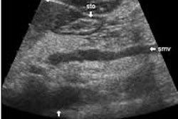
Ultrasound of both breasts was performed using a 7.5 MHz annular array transducer in both axial and sagittal planes. "Suspicious areas, such as palpable, mammographic, or sonographic abnormalities, were also scanned in a radial and antiradial orientation with and without compression, the author stated. The ultrasound criteria used to determine malignancy included ill defined or irregular borders; noncalcified lesions with posterior acoustic shadowing; architectural distortions; and solid intracystic or intraductal lesions.
3-D T2-weighted and 3-D contrast-enhanced (gadopentetate dimeglumine) T1-weighted MR imaging was performed using a 1.0-tesla scanner. A double breast coil was used with the patient in the prone position.
:We used the manufacturer's software to subtract the unenhanced images from the first and third contrast-enhanced studies in order to visualize both early- and late-enhancing lesions," they said. MR imaging findings for a malignant lesion included a focal signal that enhanced 100% or more in the first or second contrast-enhanced T2-weighted MR study or had washout. Enhancing lesions that had spiculated or irregular borders also were considered malignant.
According to the authors, if a combination of mammography and whole-breast ultrasound was performed, MRI was of no further benefit to patient management in all but seven cases (93%). Additionally, there were eight MRI studies that provided a false-positive result.
"Relevant additional findings were significantly more frequent in patients with dense breasts," they reported. "Although MR imaging is most sensitive for the detection of small tumors, routine preoperative MR imaging appears to be unnecessary for most patients if a combination of mammography and whole-breast sonography is used."
Preoperative assessment of breast cancer: sonography versus MR imagingHlawatsch, A et al
Department of radiology, Johannes Gutenberg University Hospital, Mainz, Germany
AJR 2002 December; 179:1493-1501
By Ultrasound Review
December 23, 2002
Copyright © 2002 AuntMinnie.com