(Ultrasound Review) Physicians at the University of Düsseldorf recently studied 67 patients with focal hepatic lesions, to asses whether contrast-enhanced ultrasound imaging was able to differentiate between benign and malignant lesions. While grayscale sonography is accurate in detecting focal liver lesions, its ability to separate benign from malignant is limited.
Levovist, which contains galactose and palmitic acid particles, provides a specific late phase of enhancement suitable for imaging the liver and spleen. According to the authors, "the accumulation of Levovist in the parenchyma of the liver and spleen can be made visible on sonography using the pulse-inversion mode." Their findings were reported in the November issue of American Journal of Roentgenology.
Other diagnostic modalities including liver biopsy, CT, MR imaging, or scintigraphy were used to establish that 60% of lesions were malignant, and 40% were benign. Malignant lesions included hepatocellular carcinoma, cholangiocellular carcinoma, metastases, and liver infiltration by non-Hodgkin's lymphoma. Benign lesions included focal nodular hyperplasia, hepatic adenoma, focal hyposteatosis or hypersteatosis, hemangioma, and regenerative cirrhotic nodule.
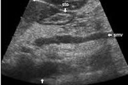
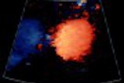
Pulse-inversion contrast imaging uses two pulses that are 180° out of phase. The summation of the two returning signals results in the elimination of linear echoes from the tissue and an image that only shows nonlinear echoes from the contrast agent. Normal liver tissue demonstrates a bright and homogeneous Levovist enhancement and focal lesions show no enhancement.
Compared with normal grayscale imaging, using an ultrasound contrast agent increased sensitivity from 85% to 100% and specificity from 30% to 63%. Also they found that, "a lower interobserver variability was found for contrast-enhanced sonography (weighted = 0.947), as compared with baseline sonography (weighted = 0.469)."
According to the authors, "all lesions that had homogeneous enhancement in the late phase of Levovist enhancement were benign. In distinction, 90% of lesions without contrast enhancement in the late phase were malignant. All lesions were malignant that were isoechoic (invisible) on baseline sonography but visible because of lack of enhancement after injection." Five additional lesions were shown after contrast enhancement that had not initially been shown.
These researchers used a 5-point scale to evaluate enhancement characteristics, with a score of one being definitely benign and five being definitely malignant. In addition, they categorized lesion using the following: homogeneous bright enhancement; dark nodules without enhancement surrounded by bright enhancement of the liver; and inhomogeneous enhancement.
They concluded, "contrast-enhanced sonography has greater specificity and sensitivity than baseline sonography for the differentiation of benign and malignant liver lesions." They demonstrated that all patients with homogeneous contrast enhancement in the lesion had benign lesions, and suggested that this form of diagnosis could replace other more invasive and expensive techniques.
Late-phase pulse-inversion sonography using the contrast agent Levovist: differentiation between benign and malignant focal lesions of the liverVon Herbay, A et al
Department of medicine and department of hepatology, gastroenterology and infectious diseases, University of Düsseldorf, Düsseldorf, Germany
AJR 2002 November; 179:1273-1279
By Ultrasound Review
December 24, 2002
Copyright © 2002 AuntMinnie.com