SAN FRANCISCO - The all-too-brief window during which stroke patients can be given brain-sparing tissue plasminogen activator (tPA) -- generally three hours -- may be extended if those patients undergo a limited MRI scan upon presentation.
By obtaining a quicker MRI exam to check for signs that intravenous tPA thrombolysis should be used, doctors in the Stroke Unit at Pitié-Salpétrière Hospital in Paris were able to administer the therapy as late as five hours after stroke onset.
And, although their patients arrived in worse condition overall than those seen in major trials of CT-guided tPA administration, the French group's MRI-guided approach achieved better survival outcomes.
The results were greeted as "good news" by an audience member who spoke after Dr. Sophie Crozier presented her group's results Thursday at the American Academy of Neurology meeting.
Previous data had suggested that screening patients with MR -- specifically, diffusion-weighted imaging (DWI) and perfusion-weighted imaging (PWI) sequences -- could extend the tPA window up to six hours, the French researchers noted.
But routine use of emergency DWI-PWI is unfeasible for most institutions because of the gadolinium injection and the difficult interpretation involved in such scanning.
"We hypothesized that a more simple MRI procedure based on DWI, FLAIR, and a short sequence of intracranial MR angiography (MRA) would provide sufficient key information to preserve the benefit/risk ratio of thrombolysis in a five-hour time-window," wrote the French researchers in their abstract.
Crozier reported the outcomes for 100 consecutive patients with middle cerebral artery (MCA) infarctions who were treated with IV tPA within a five-hour window.
Ninety-eight of the patients received the shortened DWI-FLAIR-MRA exam developed by the researchers; two patients had contraindications that precluded use of MR.
None of the patients received a CT scan, which would be standard procedure in most facilities even though CT offers less useful information on stroke patients than MR. Crozier noted that her hospital's stroke unit is able to send patients immediately to an MR scanner dedicated to neurology use.
The unit's MRI criteria for going ahead with tPA thrombolysis were:
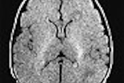
- A DWI/FLAIR pattern typical of very recent MCA infarct (DWI hypersignal without clear-cut FLAIR hypersignal); and
- Persistence of an arterial occlusion visible on MRA.
The median time between the patients' stroke onset and tPA administration was three hours and 10 minutes.
To gauge the efficacy of their approach, the researchers recorded each patient's score on the National Institute of Health Stroke Scale (NIHSS) upon admission, at 24 hours after the stroke, and seven days post-stroke. In addition, the patients' Rankin scores were measured at a three-month follow-up.
Overall, the patients were relatively young, ranging from 19-91 years old with a median age of 52. They also generally had severe strokes, with a median NIHSS score of 17. Sixty-eight of the patients had an MCA occlusion, and 30 had an intracranial ICA occlusion.
Seven patients had hemorrhages resulting from their thrombolytic therapy -- a rate comparable to that seen in large studies of CT-guided tPA administration. The researchers found no clinical or imaging predictors for the hemorrhagic response, as there was no correlation of that response with clinical severity, delay, DWI volume, or ADC values.
Arterial recanalization was achieved in 67% of the patients overall, including 70% of those with an MCA occlusion and 48% of those with an ICA occlusion. Successful recanalization was correlated with good outcomes at subsequent measurements. At three month follow-up, a Rankin score (RS) of 0-1 was achieved in 23% of the patients, RS 0-2 in 41%, RS 0-3 in 77%.
The efficacy of their MRI-based thrombolysis, as judged by 0-1 or 0-2 Rankin scores, was similar to studies of patients with similar initial severity, such as the intra-arterial PROACT2 study, the authors noted.
However, overall mortality in the study group at three months was just 8% -- much lower than the death rate reported in larger trials of CT-guided therapy, even though the French patients presented with higher NIHSS scores.
"This suggests that a simplified DWI/FLAIR and MRA MRI procedure, which is easy to implement in (clinical) routine, may contribute to extending the IV tPA time-window up to five hours," the researchers concluded.
By Tracie L. ThompsonAuntMinnie.com staff writer
April 30, 2004
Related Reading
AHA releases new guidelines for urgent stroke evaluation and management, April 4, 2003
MRI alters course of thrombolytic treatment for stroke, March 25, 2003
Perfusion CT offers speedy access, but MRI gives the "big" picture, February 19, 2003CT aids in tPA triage for stroke patients, February 17, 2003
Ministrokes may mean less damage for stroke patients, March 11, 2002
Copyright © 2004 AuntMinnie.com