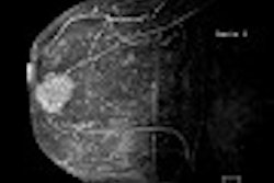
An MR technique that combines high-resolution T2-weighted and dynamic T1-weighted images is highly selective for prostate cancer staging, according to researchers from the University of Vienna in Austria.
The combined approach has a positive predictive value (PPV) exceeding 83%, said lead author Dr. Bob Djavan during a presentation at the 2003 American Urological Association meeting in Chicago. Djavan, who is the vice chair and a professor of urology at the university, also noted that the protocol addresses a need in the staging of urologic malignancy.
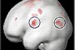
"Imaging has always been a problem in urology and prostate cancer," Djavan said. "Cancer has both neovascularization and hypoxia, and the perfusion pattern...is completely different from normal and benign tissue." The combined approach looks for patterns of neovascularization and hypoxia, he said. The study was a collaboration between investigators at the University of Vienna and the Weizman Institute in Tel Aviv.
The investigators obtained MRI images of the prostate on a 1.5-tesla unit (Magnetom Vision, Siemens Medical Solutions, Erlangen, Germany) with combined surface and endorectal coils (Prostate-Coil, Medrad, Indianola, PA). The trial involved 25 patients whose prostates were imaged prior to their undergoing radical prostatovesiculectomy (RPE).
Djavan and colleagues obtained sequences with a T2-weighted turbo spin-echo (TSE) sequence (4000/83; 5 minutes, 24 seconds; FOV 120) and dynamic Gd-DTPA-enhanced (Magnevist, Schering, Berlin) T1-weighted 3-D FLASH (8.1/4; 1 min 35 s; FOV 160; 2 pre-, 5 post-contrast acquisitions).
The researchers analyzed the dynamic T1-weighted images with a 6-time-point and 3-time-point (3TP) pharmacokinetic model. Two independent readers evaluated the T2 and T1 images. They then correlated the MRI data with histopathologic results.
The histopathologically determined tumor stage was classified according to tumor size, lymph node involvement, and the status of metastasis (TNM). Using the T2-weighted images, the readers correctly assessed the TNM stage in 21 of 25 cases (8%). In two cases (8%), tumors were understaged. In two cases (8%), tumors were overstaged.
When they used the parametrically analyzed dynamic T1-weighted images, the readers staged the tumors accurately in 19 cases (76%). In six cases (24%), tumors were overstaged.
The combined approach allowed the readers to accurately stage the tumors in 23 cases (92%). In two cases (8%), tumors were overstaged. In addition, only the combined approach allowed a correct diagnosis of extracapsular disease in all 10 cases with that level of regional spread (100%), including five cases of seminal vesicle infiltration.
Commenting on the clinical applicability of this combined approach was Dr. J. Brantley Thrasher, chair of urology at the University of Kansas Medical Center in Kansas City.
"Perfusion-enhanced MRIs have been used in more than one study," Thrasher said. "The problem is that after the biopsy has been done, we have blood that can accumulate inside or around the prostate, and this throws off the sensitivity of the test. A hematoma can appear to be cancer."
Ideally, an imaging modality would have the sensitivity to tell the physician whether surgery would be feasible due to tumor spread and other features. "We don’t have any modality sensitive enough to tell people ‘you’re not curable,’" Brantley said. "This study has a very good sensitivity, but it would have to be confirmed by more than one study group."
By Paula MoyerAuntMinnie.com contributing writer
May 15, 2003
Related Reading
AmeriScan to offer MRI prostate screening, May 1, 2003
New reports address prostate cancer staging, therapy, March 24, 2003
Predictors of PSA relapse after radiotherapy for prostate cancer identified, May 17, 2003
Older age predicts shorter survival with high-risk prostate cancer, January 28, 2003
Copyright © 2003 AuntMinnie.com