Although FDG-PET is considered the gold standard for assessing myocardial viability, late-enhancement (LE) MRI may offer a reasonable alternative, according to German researchers.
In addition to comparing LE-MRI imaging with PET, Dr. Hubertus Bülow and colleagues from the Technischen Universität in Berlin evaluated the intra- and interobserver variability and reproducibility of LE in MRI. They discussed their results at the 2002 Society of Nuclear Medicine meeting in Los Angeles.
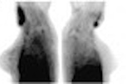
The study included 10 patients with stable coronary artery disease and significantly reduced left ventricular function. Each patient was scanned once with FDG-PET, and twice with MRI over a 5-12 month interval, following the administration of gadolinium (III) diethyltriaminepentaacetic (Gd-DTPA). No cardiac events or revascularization procedures occurred between MRI scans.
MRI was performed using a gradient-echo technique with echo-planar readout 20 minutes after intravenous administration of 0.2 mmol/kg body weight Gd-DTPA. FDG uptake and contrast enhancement were compared and analyzed for intraobserver and interobserver variability, as well as MRI reproducibility. Intraobserver was defined as 1 scan, 1 observer, and 2 readouts at a time interval of 3 minutes. Interobserver is defined as 1 scan and 2 independent observers.
According to the results, assessment of myocardial viability in MRI with the LE after Gd-DPTA application is a method comparable to FDG-PET, the authors wrote. Analysis of LE contrast enhancement on a segmental basis yielded a high concordance for intra- (c 2=370, p<0.001) and interobserver (c 2=461, p<0.001) investigation. In addition to low intra- and interobserver variability, segmental analysis of the CE LE MRI technique demonstrates a high reproducibility in left ventricular localization and the degree of transmural involvement over a time period of about one year (c 2=254, p<0.001).
"LE-MRI may be an alternative to FDG-PET for imaging scar tissues, chronic myocardial infarction, and left ventricular dysfunction, but not hibernating myocardial (tissue)," Bülow said. The modality seems particularly adept at detecting tissue that has irreversible damage, he added. Future studies will have to look at whether LE MRI offers any cost benefit over FDG-PET.
By Laura Ruth
AuntMinnie.com contributing writer
August 26, 2002
Related Reading
Dobutamine SPECT helps assess myocardial viability, August 19, 2002
Low-dose dobutamine enhances SPECT detection of myocardial viability, June 19, 2002
Noninvasive MCE fares slightly better than MRI for tracking cardiac perfusion defects, March 8, 2001
Copyright © 2002 AuntMinnie.com