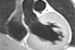
DENVER - Proton MR spectroscopy may offer a clearer view of the size and borders of untreated gliomas than conventional imaging, researchers reported at the American Academy of Neurology meeting on Monday.
"These results support the conclusion that proton magnetic resonance spectroscopy accurately reflects the glioma tumor burden," said Dr. David Croteau of the Henry Ford Hospital in Detroit. "This has important diagnostic and therapeutic implications for the accurate assessment of burden of disease in glioma patients."
Croteau and his colleagues compared proton MR spectroscopy with image-guided biopsies, using semiquantitative and qualitative histopathologic analyses, from a series of 39 untreated patients whose tumors produced 313 tissue samples. A 1.5-tesla MR scanner was used, along with a multivoxel acquisition technique. Regions of interest were referenced to the same spatial coordinates.
Gliomas are a particularly difficult tumor to treat because of areas of necrosis, solid tumors, and varying degrees of tumor infiltration into normal tissue, Croteau said. However, proton MR spectroscopy allows for the identification of the degree of infiltration. A metabolic ratio, using choline-containing compounds as the numerator and creatine or N-acetyl-aspartate as the denominator, appears to correlate with degree of infiltration.
The aim of the study was to use proton MR spectroscopy to better define the microscopic anatomy of glioma on imaging studies. The system has been used previously to improve the characterization of tumors and the surrounding brain tissue, he said.
"Despite the interpatient overlap of metabolic ratios between normal tissue and mild tumor infiltration," preliminary analyses revealed that proton MR spectroscopy appears more accurate than conventional MR imaging in delineating the tumor boundary and quantifying the degree of tumor infiltration, Croteau said.
"These ratios appear to be a useful tool which may help define selection of biopsy, enhance and define the size of gliomas, assess residual disease, and monitor the effects of surgery and chemotherapy in order to optimize treatment," he concluded.
By Edward SusmanAuntMinnie.com contributing writer
April 16, 2002
Related Reading
Watchful waiting may suffice for patients with suspected low-grade glioma, March 23, 2001
MRI and SPECT garner high marks for glioma grading, March 5, 2001
MR perfusion helps diagnose brain lesions, February 16, 2001
Copyright © 2002 AuntMinnie.com