According to FDG-PET results, patients who have recovered from depression using Cognitive Behavioral Therapy (CBT) experience different brain-pattern changes compared with patients who have recovered using drugs, Canadian researchers report in the Archives of General Psychiatry.
"Our findings set the stage for future research to identify imaging markers in individual subjects, with hopes to discern evidence-based imaging markers for the optimization of treatment protocols," said principal investigator Dr. Helen Mayberg, a professor of psychiatry and neurology at Emory University in Atlanta.
At the time of the study, Mayberg was a senior scientist at The Rotman Research Institute at Baycrest Center for Geriatric Care in Toronto, and professor of neuropsychiatry at the University of Toronto. The research was conducted at Rotman, the university and Toronto’s Center for Addiction and Mental Health.
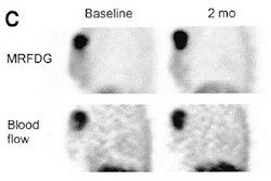
The researchers studied brain changes underlying the response to CBT by using resting-state 18FFDG-PET. They scanned 17 unmedicated, unipolar depressed outpatient subjects (mean age 41 at enrollment) before and after 15-20 sessions of CBT. They used whole-brain, voxel-based methods to evaluate response-specific CBT effects.
They made a post hoc comparison to an independent group of 13 paroxetine-treated, treatment responsive patients to identify and compare the specificity of action of the two treatments on the brain.
"A full course of CBT resulted in significant clinical improvement in the 14 study completers," the researchers reported. "Treatment response was associated with significant metabolic changes: increases in hippocampus and dorsal cingulated (Brodmann area - BA-24) and decreases in dorsal (BA 9/46), ventral (BA 47/11) and medial (BA9/10/11) frontal cortex. This pattern is distinct from that seen with paroxetine-facilitated clinical recovery, where prefrontal increases and hippocampus and subgenual cingulate decreases were seen" (Archives of General Psychiatry, January 2004, Vol. 61:1, pp. 34-41).
Based on these findings and further research, PET scans could eventually become a significant component in the treatment and monitoring of clinical depression, Mayberg suggested.
"One way of treating patients clearly does not fit everyone," she added. "This imaging study shows us in a new way that depression can be treated effectively by a variety of pathways."
By Bruce SylvesterAuntMinnie.com contributing writer
January 7, 2004
Related Reading
Prolonged depression reduces global cerebral gray matter volume, November 20, 2003
Illuminating the mind: Imaging explores the mysteries of meditation, September 11, 2003
MR study shows antidepressants shield hippocampus from deterioration, August 5, 2003
Copyright © 2004 AuntMinnie.com