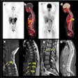
VIENNA - Fusion PET-CT scanners offer a powerful combination of functional and anatomical data that can give radiologists precise information on tumor location and stage. But MRI still has a strong role to play in oncology imaging, as both modalities demonstrate different strengths and weaknesses in tumor visualization, according to German researchers at this week's European Congress of Radiology.
A group from University Hospital in Essen presented a comparison of PET-CT and MRI in Monday's ECR scientific sessions. The group also discussed its work in using PET-CT to track radiation therapy treatment, as well as in reducing respiratory artifacts on PET-CT studies with a new breathing protocol.
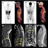
In the first study, Dr. Gerald Antoch compared PET-CT to MRI in visualizing tumors in 50 patients. The group used a dual-slice biograph PET-CT scanner (Siemens Medical Solutions, Erlangen, Germany) and a 1.5-tesla Magnetom Sonata MRI unit (Siemens), equipped with a rolling table (bodySurf). The field of view was set from the head to the upper thigh, and patients received MRI scans with a gadolinium-based contrast agent.
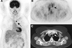
The modalities were found to be comparable in 33 (66%) patients, and the abdominal organs were assessed equally well in all patients, Antoch said. PET-CT beat MRI in detecting lymph node and pulmonary lesions, however.
In eight patients (16%) with cervical, thoracic, or abdominal lymph node tumors, PET-CT was able to detect tumors missed on MRI due to their small size (less than 1 cm). PET-CT spotted 94 pulmonary lesions in 16 patients, compared to 58 lesions in 13 patients for MRI.
MRI excelled in visualizing bone metastases, however, finding metastases in 7 patients (14%) that were missed on PET-CT.
"Whole-body PET-CT seems to be superior to whole-body MRI in detecting lymph node metastases and pulmonary lesions," Antoch said. "On the other hand, whole-body MRI detects bone metastases more accurately. Whole-body PET-CT and MR imaging complement each other in the staging of malignant diseases."
PET and radiation therapy
Antoch went on to present the next study, in which the Essen group examined the utility of PET-CT to monitor radiation therapy of liver tissue in a pig model. Nine pigs underwent central bile duct resection, followed by biliodigestive anastomosis.
Six of these pigs were treated with 20 Gy of radiation therapy to the area of the anastomosis. PET-CT liver scans were then conducted at two, four, and eight weeks to assess the impact of the irradiation on liver function as measured by glucose uptake.
The follow-up scans found sharply decreased glucose utilization in the irradiated area at two weeks, with glucose uptake increasing over time until there was no difference in uptake between the irradiated area and the rest of the liver at eight weeks. Meanwhile, CT scans were able to demonstrate an increase in liver volume and a bile duct diameter significantly higher compared to the pigs that didn't receive radiation therapy.
Antoch concluded that PET is well suited for delineation of functional alterations following radiation therapy, while CT is appropriate for visualizing surgical alterations, vascular differences, and liver volume. Antoch recommended first assessing an irradiated area with PET-CT, and then following up with PET and CT independently.
Finally, another Essen researcher, Dr. Thomas Beyer, explained the group's development of a new breathing protocol designed to reduce the impact of respiration-induced motion artifacts during PET-CT studies with single-slice scanning. Such artifacts can cause misalignment between the PET and CT images, he said.
The group separated 80 patients into three groups: 20 were scanned with a tidal breathing technique in single-slice mode, 30 with tidal breathing in dual-slice mode, and 30 in single-slice mode using a protocol that the group developed. The Essen group recently upgraded its biograph system from a single-slice CT to a dual-slice model, giving the group the ability to compare both modes.
The technique asked patients to limit their breathing during crucial parts of the image acquisition cycle. The patient begins by breathing quietly, and as the scan's field of view reaches the mediastinum the patient is told to exhale and hold their breath for next 10-15 seconds, the amount of time it takes for the scanning slice plane to reach the lower end of the liver. The patient is then allowed to continue breathing again while the CT scan collects data in the lower pelvis.
The group found that, without using the protocol, there were displacements of up to 25 mm between the CT and PET images in the diaphragm area (the zone most effected by respiration-related movement). Because CT data were also used for attenuation correction, the artifacts created the impression of FDG uptake in the lung and liver.
There were 25% more artifact-free cases when using the breath-hold protocol in single-slice mode, and 50% more artifact-free cases when the biograph was used in dual-slice CT mode. Beyer said that good patient preparation in advance of the limited breath-hold technique is key to ensuring a scan with few serious artifacts.
By Brian CaseyAuntMinnie.com staff writer
March 10, 2003
Related Reading
Physician groups navigate a rocky road to PET reimbursement, February 26, 2003
PET useful for staging pediatric bone sarcomas, February 7, 2003
Studies illustrate benefits of FDG-PET, pre-planning in radiotherapy, January 27, 2003
PET/CT preferred as whole-body scan for cancer detection, December 6, 2002
Combined PET and MRI distinguish necrosis from tumor recurrence, October 29, 2002
Copyright © 2003 AuntMinnie.com

