The radiation exposure to staff and patients during three types of CT-guided procedures -- coagulation of osteoid osteoma, biopsy, and drainage -- is well within acceptable ranges, according to a study in The British Journal of Radiology.
Researchers from Leiden University Medical Center in the Netherlands retrospectively assessed the radiation dose received from repeat CT scans at the same anatomical locus during image-guided interventional procedures.
"There is concern about the occurrence of skin damage when threshold dose levels for deterministic skin effects, such as erythema or epilation, are exceeded," the authors stated (BJR, August 2001, Vol. 74:885, pp. 720-726).
The imaging exams reviewed for this study were performed on a conventional Tomoscan LX CT scanner. The CT fluoroscopic procedures were done on a Tomoscan AV-E1 spiral CT scanner (Philips Medical Systems, Bothell, WA). During fluoroscopy, the CT image is refreshed 6 times per second with a minimum fluoroscopy duration of 1 second. All CT fluoroscopy was performed with the quick-check method so that the radiologist’s hands were not exposed to significant radiation, the authors wrote.
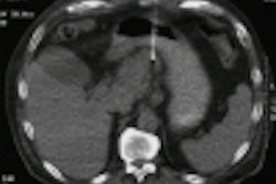
Patient dose and maximum entrance skin dosage (ESD) was assessed for 96 CT-guided interventions, including 26 drainages, 49 biopsies, and 31 coagulations of osteoid osteoma.
"Dose measurements were performed to derive the [computed tomography dose index, CTDI] at both the Philips LX and the AV-E1 scanner. This included measurements at all available tube potentials and slice thicknesses. Personnel dosimetry was performed with electronic personal dosimeters," they wrote.
According to the results, on the LX scanner, the localizing scan preceding the puncture was most often a contiguous scan at a tube voltage of 120 kVp and a slice thickness of 10 mm, for drainage or biopsy, or 3 mm for coagulation of osteoid osteoma.
CT fluoroscopy slices during drainages and biopsies were collected at 140 kV tube voltage for the abdomen and 120 kV for the chest, both at 25 mAs per rotation tube load and 3-7 mm slice thicknesses.
"The maximum measured dose to one worker during one procedure was 28 µSv. Average doses per procedure to the radiologists are well below 10 µSV, and average doses to the radiographers are below 1 µSv per procedure," they stated.
A skin dose of 1 Gy will be reached only after CT fluoroscopy was performed for 2.5 to 8 minutes at the same location, which is "much longer than the CT fluoroscopy times observed at our CT scanner."
In addition, the ESD was below the 2 Gy threshold for deterministic skin doses on a CT scanner equipped with fluoroscopic function.
One reason for the lower fluoroscopic dosage was the quick-check method itself, which requires only a single shot of fluoroscopy after needle manipulations, the authors concluded. In the end, the cumulative annual dose due to CT fluoroscopy to the single radiologist who performed about 70% of the procedures for this study was estimated at less than 0.1 µSv.
By Shalmali PalAuntMinnie.com staff writer
October 5, 2001
Related Reading
Low-dose CT fluoroscopy minimizes interventional exposure, July 30, 2001
AJR review chronicles fluoroscopy-induced skin damage, June 21, 2001
New generation of digital fluoro systems can reduce radiation exposure, March 3, 2001
Copyright © 2001 AuntMinnie.com