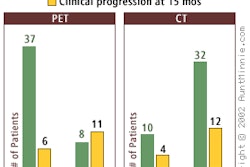
A multinational research team has successfully imaged amyloid plaques in 14 Alzheimer’s patients using PET and a compound developed at the University of Pittsburgh School of Medicine. Amyloid plaques are one of the pathological hallmarks of Alzheimer's disease.
The results of these studies were presented at International Conference on Alzheimer's Disease and Related Disorders this week in Stockholm.
More than 100 compounds were analyzed over a ten-year period before a team, led by Chester Mathis, Ph.D., selected the compound 6-OH-BTA-1 because of its ability to bind specifically to amyloid plaques, cross the blood brain barrier, and clear from normal brain tissue.
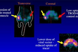
The Pittsburgh team, along with Drs. Brian Bacskai and Brad Hyman at Massachusetts General Hospital in Boston, tested the compound on transgenic mice with Alzheimer’s disease. Using microscopic multiphoton imaging, researchers were able to inject the compound into the mice and see individual amyloid plaques as they came in contact with the 6-OH-BTA-1 compound.
Human testing was conducted at the Uppsala University PET Center in Uppsala, Sweden by researchers from the Karolinska Institutet, in Huddinge, Sweden. The group was lead by Bengt Langstrom, a professor in organic chemistry at the university. Nine of the 14 subjects had been diagnosed with Alzheimer’s; the remaining 5 subjects were a healthy control group. After receiving the compound intravenously, the study participants underwent a 60-minute PET scan.
Images showed that the compound was retained in brain regions consistent with the accumulation of plaques. Previously, amyloid plaques have only been seen during autopsy in the brains of people with Alzheimer’s disease. In addition, the researchers found that little of the compound was retained in the brains of the control group.
The compound, also known as Pittsburgh Compound B (PIB), shows promise in the development of new treatments for Alzheimer’s.
"Having the capability to quantify amyloid deposition in the brain will have a profound impact on our ability to monitor the progression of Alzheimer’s as well as gauge the effectiveness of medical treatments," said William Thies, Ph.D., vice president of medical and scientific affairs for the Chicago-based Alzheimer’s Association.
By Jonathan S. BatchelorAuntMinnie.com staff writer
July 23, 2002
Related Reading
New tracers, technologies propel clinical applications in PET, June 14, 2002
CMS panel dashes hopes for PET Alzheimer’s payments, April 22, 2002
U.S. gives thumbs-down on PET for Alzheimer's, January 11, 2002
CMS tackles FDG-PET reimbursement for Alzheimer’s, January 10, 2002
PET scanning plus new molecular probe detects early Alzheimer's disease in vivo, January 10, 2002
Copyright © 2002 AuntMinnie.com