Preoperative ultrasound categorizes most borderline ovarian tumors as intermediate and high-risk (O-RADS 4 and 5), suggest findings presented at ARRS 2026 in Pittsburgh.
In her talk, Erika Baca, MD, from the University of New Mexico in Albuquerque discussed her team’s findings, which suggest that O-RADS appropriately stratifies risk for these adnexal lesions.
“This is clinically important as the treatment for borderline ovarian tumors may or may not spare fertility, and these tend to occur in younger patients,” Baca said.
Borderline ovarian tumors are of epithelial origin and come in serous and mucinous subtypes. While the 10-year survival rate exceeds 95%, about one-third of cases occur in women younger than 40. Fertility-preserving treatment may be considered, but if there are no fertility requirements, then total hysterectomy and bilateral salpingo-oophorectomy are recommended.
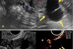
Baca and colleagues evaluated the distribution and determined risk factors of pathologically-proven borderline ovarian tumors. They used the 2022 version of the O-RADS lexicon to help determine how preoperative ultrasound imaging can best be used for early and accurate diagnosis of these tumors.
The study included 60 women with an age range of 18 to 89 years. The women had an adnexal mass with pathologically confirmed borderline ovarian tumor diagnosis. They underwent pelvic ultrasound between 2020 and 2023 within 12 months prior to the adnexal mass surgery. Independent evaluators assigned O-RADS scores based on ultrasound features collected per the O-RADS lexicon.
All but one case had either a designated O-RADS 4 (n = 36) or 5 (n = 23) category. The one exception had an O-RADS 2 designation. Of the intermediate and high-risk tumors, 42 were serous while 17 were mucinous.
Using univariable and multivariable analyses of ultrasound imaging features, Baca et al reported color score as a strong risk factor. While external contour was a strong but statistically insignificant factor on univariable analysis, it proved to be a weak risk factor on multivariable analysis.
Among other findings, most borderline ovarian tumors showed minimal to moderate flow, had smooth contouring, and were non-solid. In addition, 33 tumors were 10 cm in diameter or greater while 26 were smaller than 10 cm.
With intralesional vascularity being the strongest O-RADS lexicon descriptor and predictor of borderline ovarian tumors, Baca said this can be tricky.
“It can argued as the most subjective [predictor] with how people are grading how much flow is going to the lesion,” she said.
Baca said larger sample sizes could help validate the team’s findings and eventually apply this approach to the MRI O-RADS lexicon.

![Examples of ultrasound findings and techniques. (A) Images in a 39-year-old male patient with a mass in the left thigh. The mass is heterogeneous on the B-mode US image (compared with the patient in D) and showed increased microvascularity (superb microvascular imaging [SMI]) and shear-wave elastography (SWE) values. Undifferentiated pleomorphic sarcoma was diagnosed at biopsy (with pleomorphic rhabdomyosarcoma in surgical specimen). (B) Images in an 18-year-old male patient with a mass in the left leg. The mass is hypoechoic on the B-mode image, with no other findings suggestive of malignancy. The lesion is in contact with the cortex of the tibia, which is slightly irregular. CT revealed a doubtful anteromedial tibial erosion. The microvascular study demonstrated high vascularization, suggestive of malignancy. Periosteal Ewing sarcoma was diagnosed with both histologic and immunohistochemical confirmation. (C) Images in a 69-year-old female patient with a lump growing on the outside of the left leg. Multiple SWE examinations were performed (please note the high values obtained in the measurements, whereas the color map highlights the stiffness relative to adjacent tissues). SMI showed areas of increased vascularization to target for sampling. Undifferentiated spindle cell sarcoma was diagnosed at biopsy, with residual leiomyosarcoma in the surgical specimen after neoadjuvant therapy. (D) Images in a 56-year-old female patient with a mass in the right thigh. The mass is heterogeneous at both B-mode ultrasound (similar to patient A) and MRI (coronal T2-weighted spectral attenuated inversion recovery [SPAIR]; T1-weighted pre-contrast and postcontrast imaging), which even shows uptake after the administration of paramagnetic contrast material, which is traditionally suggestive of malignancy. Low values at SMI and elastography are suggestive of benignity. Spindle cell lipoma was diagnosed at biopsy, with atypical spindle cell lipomatous tumor in the surgical specimen.](https://img.auntminnie.com/mindful/smg/workspaces/default/uploads/2026/08/images-radiol250278fig2.APCFLSvX6p.jpg?auto=format%2Ccompress&fit=crop&h=100&q=70&w=100)


![Examples of ultrasound findings and techniques. (A) Images in a 39-year-old male patient with a mass in the left thigh. The mass is heterogeneous on the B-mode US image (compared with the patient in D) and showed increased microvascularity (superb microvascular imaging [SMI]) and shear-wave elastography (SWE) values. Undifferentiated pleomorphic sarcoma was diagnosed at biopsy (with pleomorphic rhabdomyosarcoma in surgical specimen). (B) Images in an 18-year-old male patient with a mass in the left leg. The mass is hypoechoic on the B-mode image, with no other findings suggestive of malignancy. The lesion is in contact with the cortex of the tibia, which is slightly irregular. CT revealed a doubtful anteromedial tibial erosion. The microvascular study demonstrated high vascularization, suggestive of malignancy. Periosteal Ewing sarcoma was diagnosed with both histologic and immunohistochemical confirmation. (C) Images in a 69-year-old female patient with a lump growing on the outside of the left leg. Multiple SWE examinations were performed (please note the high values obtained in the measurements, whereas the color map highlights the stiffness relative to adjacent tissues). SMI showed areas of increased vascularization to target for sampling. Undifferentiated spindle cell sarcoma was diagnosed at biopsy, with residual leiomyosarcoma in the surgical specimen after neoadjuvant therapy. (D) Images in a 56-year-old female patient with a mass in the right thigh. The mass is heterogeneous at both B-mode ultrasound (similar to patient A) and MRI (coronal T2-weighted spectral attenuated inversion recovery [SPAIR]; T1-weighted pre-contrast and postcontrast imaging), which even shows uptake after the administration of paramagnetic contrast material, which is traditionally suggestive of malignancy. Low values at SMI and elastography are suggestive of benignity. Spindle cell lipoma was diagnosed at biopsy, with atypical spindle cell lipomatous tumor in the surgical specimen.](https://img.auntminnie.com/mindful/smg/workspaces/default/uploads/2026/08/images-radiol250278fig2.APCFLSvX6p.jpg?auto=format%2Ccompress&fit=crop&h=112&q=70&w=112)