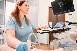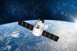
ORLANDO -- Sonographers on Earth can take a page or two from astronauts who use ultrasound in space, according to presentations given at the 2025 American Institute of Ultrasound in Medicine (AIUM) annual convention.
In their talks, Leroy Chiao, PhD, and Scott Dulchavsky, MD, PhD, from Henry Ford Health in Detroit, Michigan discussed ultrasound’s utility in micro-gravity environments such as aboard the International Space Station (ISS). They also presented on how ultrasound research has helped improve the health of astronauts in such environments and outcomes for patients on the ground.
“Ultrasound is really pushing the boundaries of what’s possible in using a relatively low-cost, low-weight machine compared to a CT scanner or an MR machine,” Chiao said. “Ultrasound has really helped space medicine quite a bit and will continue to expand.”
Ultrasound is the only imaging modality currently used in space, with point-of-care ultrasound (POCUS) tools examining astronauts for various conditions. These include muscle degeneration, exposure to radiation, spinal conditions, orthostatic tolerance, and blood volume decreases, among others.
Chiao, a former NASA astronaut and ISS commander, was part of the Advanced Diagnostic Ultrasound in Microgravity (ADUM) project. This project investigated strategies for applying diagnostic telemedicine to space. It was the first formal study that explored the use of ultrasound in microgravity.
Chiao told personal stories of his time in space, along with how ultrasound findings have helped in developing health measures and diagnostic protocols for spaceflight. He also explained the various effects that space travel has on the human body and how daily exercise is assigned to astronauts to combat these effects.
Leroy Chiao, PhD, discusses the effects of space travel on the human body, as well as ultrasound's utility in microgravity conditions.
Dulchavsky taught Chiao about how to use ultrasound aboard spacecraft, which the latter said was “surprisingly easy.”
“He came to Houston and trained us for just a matter of hours. It was mostly about pattern recognition, recognizing what made a good ultrasound image or a bad ultrasound image,” Chiao said. “Very quickly, we were able to become pretty good remote operators of the device.”
Dulchavsky’s presentation meanwhile focused on useful information that ultrasound provides in diagnosing the aforementioned conditions astronauts may experience while in space. He has co-authored studies showing the performance of ultrasound in microgravity environments, such as diagnosing broken bones, collapsed lungs, problems with intracranial pressure, and sinusitis.
Some advantages of ultrasound include being cost-effective, being non-radioactive, and being expansive, Dulchavsky added.
“With an ultrasound machine, you can use it for anything from problems with muscles and bones and brains,” he said. “It’s a single device that provides way more information than anything we’ve had before.”
Scott Dulchavsky, MD, PhD, talks about take-home messages from space that sonographers can use in their practice.
The remote medical capabilities that astronauts have can also be implemented for practices on Earth. For example, POCUS can be used to image patients in remote areas. Also, best practices on collaboration and promoting mental wellness in an isolated environment such as on the ISS can be used by sonography teams.
Dulchavsky added that sonographers should get creative when faced with imaging challenges, going after problems with innovation.
“If you only have one device that provides diagnostic capabilities, can you go beyond what would be normally done to solve a problem?” he said. “I hope my talk inspires people to look at other things that I haven’t even thought about. I can think with the newer techniques and technologies that are now being employed with ultrasound devices we have now…the diagnostic capabilities are limitless.”