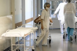
Radiology staff at the Ohmatdyt Children's Hospital in Kyiv are making steady progress with their plan to restore normal service after a devastating missile attack.
On July 9, 2024, AuntMinnieEurope.com posted an article about the extensive damage caused to Ukraine's largest pediatric hospital. Stanislav Rebenkov, MD, head of radiology, and his team are gradually starting to rebuild the department.
 Stanislav Rebenkov, MD, has been moved by the support and generosity of the global radiology community.
"On Friday, our hospital received 7 Butterfly iQ+ ultrasound devices, which will be indispensable in our work with the pediatric patients," he said in a Facebook post on October 21. "These devices will significantly enhance our diagnostic capabilities, especially in the intensive care, emergency, and pediatric departments."
The donation was the work of First Read Initiative (FRI), a charitable fund that aims to make diagnostic imaging accessible to patients in unserved and underserved regions around the globe, in collaboration with Douglas Davis, MD, and Olga Maihutiak, MD, at MedGlobal and the Ukrainian Medical Association of North America (UMANA).
 Much rebuilding work still needs to be done at the Ohmatdyt Children's Hospital.
Radiologist Josh Sokol, MD, a founder of the FRI who works with teleradiology company vRad, read AuntMinnieEurope.com's July 9 article and recognized the urgent need for ultrasound capabilities at the Ohmatdyt. He assisted with the logistics of the delivery process for the Butterfly devices.
Uliana Pidvalna, MD, associate professor in the department of normal anatomy at Danylo Halytsky at Lviv National Medical University, and musculoskeletal radiologist Volker Lapczynski, MD, from Unilabs in Oslo, Norway, played a key role in establishing the partnership between vRad/FRI and MedGlobal Ukraine. Earlier this month, Sokol attended the MedGlobal annual congress in Chicago and met with Maihutiak plus many other Ukrainian physicians.
"As a result of this conference, we will be working with some Ukrainian intensive care specialists and others to provide frontline point-of-care ultrasound to help aid in the triage and care of soldiers and victims in Ukraine," he noted.
 Clear evidence is still apparent of the damage caused by the rocket attack.
In his Facebook post, Rebenkov thanked MedGlobal and FRI -- particularly Davis, Sokol, and Maihutiak and their teams – for their support. "This project would not have been possible without you. We deeply appreciate your efforts in organizing, transporting, and personally delivering this vital equipment to Kyiv during the International Healthcare Summit held at Shupyk National Healthcare University of Ukraine on October 15-17."
Rebenkov also paid gratitude to the radiology community, which has actively supported the staff at the Ohmatdyt since the missile attack carried out by Russia.
 Thanks to the support of the global imaging community, radiology staff at the Ohmatdyt can now look forward to restoring normal service.
"Your solidarity and contributions have been invaluable during this challenging time, helping us restore operations and continue providing critical care to our patients," he noted. "Your contributions not only help us continue providing high-quality medical care but also greatly expand our capabilities in diagnosing and treating children with severe conditions. You are helping save lives and giving hope to those who need it most!"