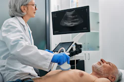
Machine learning can help improve echocardiography interpretation of heart tumors, according to research published on July 1 in Informatics in Medicine Unlocked.
A team led by Seyed-Ali Sadegh-Zadeh, PhD, from Staffordshire University in England found that its machine-learning model achieved high performance in diagnosing heart tumors, including a near-perfect area under the curve (AUC) score.
"These findings advocate for the potential of machine learning in revolutionizing cardiac tumor diagnostics, offering pathways to more accurate, noninvasive, and patient-centric diagnostic processes," the Sadegh-Zadeh team wrote.
While rare, cardiac tumors present unique challenges for clinicians due to symptoms mimicking other conditions. Localization and characterization of these tumors require advanced imaging.
Echocardiography is the primary imaging modality for this area, but its ability to differentiate between tumor types and determine malignancy is limited. The researchers highlighted that machine learning techniques could lead to improved diagnostic performance.
Sadegh-Zadeh and colleagues integrated data from echocardiography images and pathology with advanced machine-learning techniques to improve the diagnostic accuracy of cardiac tumors. They used support vector machines, random forest, and gradient boosting machines that were optimized for limited datasets in specialized medical fields.
The study included clinical data from 399 patients and evaluated the performance of the models against traditional diagnostic metrics. The researchers reported that the random forest model was superior to the other models in accurate diagnosis.
| Performance of machine-learning models in diagnosing heart tumors | |||
|---|---|---|---|
| Measure | Support vector machines | Gradient boosting machines | Random forest |
| Accuracy | 71.25% | 96.25% | 96.25% |
| Precision (benign tumors) | 78% | 99% | 99% |
| Precision (malignant tumors) | 50% | 88% | 88% |
| Recall (benign) | 43% | 95% | 95% |
| Recall (malignant) | 43% | 99% | 99% |
| F1 score (benign) | 80.34 | 97.3% | 97.3% |
| F1 score (malignant) | 46.51 | 93.88% | 93.88% |
| AUC | 0.72 | 0.98 | 0.99 |
The team also identified the following key clinical predictors: age, echo malignancy, and echo position. This underscores the value of integrating diverse data types, they noted.
The random forest model was included in clinical validation and achieved a diagnostic accuracy of 94% in a real-world setting.
The study authors highlighted that the results show machine learning's capabilities in improving diagnostic precision in assessing heart tumors. They added that the study "also sets a foundation for future explorations" into broader applications for the technology across various domains of medical diagnostics. It emphasizes the need for expanded datasets and external validation, the authors noted.
"Additionally, examining implementation studies to understand the practical aspects of integrating these models into clinical settings, including workflow integration, clinician training, and patient outcomes, is vital for successful adoption," they wrote.
The full study can be found here.