AUSTIN, TX – Point-of-care ultrasound (POCUS) isn’t typically used for imaging the pancreas, but perhaps that should change, according to a presentation given April 7 at UltraCon.
In her talk, Alice Lee, MD, from Stanford University in California highlighted findings on how POCUS can be a reliable tool for pancreatic imaging, whether performed by experienced or novice sonographers.
“One of the big implications of pancreatic POCUS is the way that we put imaging in the hands of a provider who’s really managing the patient, whether it’s the gastroenterologist or the primary care physician,” Lee said.
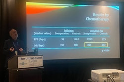
 Alice Lee, MD, from Stanford University presents research on the feasibility and performance of point-of-care ultrasound (POCUS) at UltraCon.Amerigo Allegretto
POCUS has gained traction and popularity in the 21st century for its convenience, versatility, and cost-effectiveness, but it isn’t widely used for imaging of the pancreas.
“In the U.S., we’ve kind of pooh-poohed this idea of ultrasound for the pancreas with the bile gas, obesity, and it being a retroperitoneal organ,” Lee said. “But we’ve found through colleagues in other parts of the world that there are areas where … some countries in Asia do use ultrasound for basic first-line imaging for everything, including the pancreas. There are studies that show that in doing that, they [physicians] are able to catch pancreatic cancer earlier.”
Lee suggested that operator dependency and limitations of current ultrasound technology may contribute to the lack of usage in the U.S.
In a pilot study, Lee and colleagues examined the feasibility of POCUS in pancreatic imaging. They included data from 40 patients with an average age of 61.3 years within Stanford Hospital’s benign pancreas clinic and endoscopy unit.
Two physicians operated the POCUS exams. One was a radiologist with more than five years of ultrasound experience who was blinded to clinical data. The other was a gastroenterologist with four hours of ultrasound training who was not blinded to the clinical data. Both physicians performed independent bedside POCUS exams and documented visibility and exam length. The researchers compared the findings to recent CT, MRI, and/or endoscopic ultrasound exams.
The team reported that the physicians obtained complete or partial images in most exams, although the radiologist showed superior performance. The physicians struggled with imaging the tail area of the pancreas.
| Comparison of visibility rates between radiologist, gastroenterologist in pancreatic POCUS (out of 40 images) | ||||
|---|---|---|---|---|
| Radiologist | Gastroenterologist | |||
| Pancreatic area | Complete or partial images | None | Complete or partial images | None |
| Head | 38 | 2 | 34 | 6 |
| Body | 40 | 0 | 32 | 8 |
| Tail | 27 | 13 | 15 | 25 |
Additionally, they had comparable exam times, including four minutes for the radiologist and five minutes for the gastroenterologist.
Compared with other imaging modalities, POCUS as performed by the radiologist determined normal pancreatic findings in all cases. Additionally, it accurately visualized 75% of cysts and masses 1 cm or larger and 75% of dilated pancreatic ducts as found on CT, MRI, and/or endoscopic ultrasound.
However, POCUS struggled with finding smaller cysts and masses, parenchymal atrophy, hyperechoic stranding, and lobularity.
Lee said that with these findings in mind and despite the rudimentary nature of the exams, the future of pancreatic POCUS is promising.
One way that clinics in Asia employ to help improve visualization is having patients drink room-temperature or warm distilled water to create more contrast between the pancreas and stomach, according to Lee. She added that contrast enhancement and quantitative ultrasound tissue characterization could also improve visibility through POCUS.
“We also think there are promising implications for teaching given Physician B’s (gastroenterologist) results,” she said. “Physician B did a decent job despite only four hours of treatment.”
Lee said that the next steps include exploring how liquid ingestion, contrast enhancement, and quantitative ultrasound characterization can further visualize pancreatic findings.