NYU Langone Health’s Perlmutter Cancer Center is using histotripsy for some liver cancer cases, it said in a March 28 blog post.
Histotripsy uses focused ultrasound to deliver external beams intended to destroy cancerous tissue while leaving surrounding tissue intact. Histotripsy could become an option for patients who might otherwise be unlikely candidates for radiation or other ablation techniques, according to NYU Langone Health.
"This device allows us to offer ablation to patients who would normally not be good candidates," said Mikhail Silk, MD, an interventional radiologist at NYU Langone who performed the procedure. “Even more exciting is the potential abscopal effect in patients with additional disease or microscopic tumors not caught by scans, shrinking them alongside the targeted tumors. The data on these additional benefits are preliminary, but it is a promising avenue for further research.”
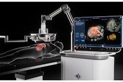
Mikhail Silk, MD, an interventional radiologist at NYU Langone Health, leads a mock session in the setup of focused ultrasound histotripsy procedures now available through Perlmutter Cancer Center in New York City. Photo courtesy of NYU Langone Health.
NYU Langone is also testing the HistoSonics Edison System as part of the HOPE4KIDNEY US clinical trial for the treatment of kidney tumors. Led by William Huang, MD, the trial is expected to last about seven years, including an enrollment phase of around 18 months and follow-up for up to five years after the procedure.








![Examples of ultrasound findings and techniques. (A) Images in a 39-year-old male patient with a mass in the left thigh. The mass is heterogeneous on the B-mode US image (compared with the patient in D) and showed increased microvascularity (superb microvascular imaging [SMI]) and shear-wave elastography (SWE) values. Undifferentiated pleomorphic sarcoma was diagnosed at biopsy (with pleomorphic rhabdomyosarcoma in surgical specimen). (B) Images in an 18-year-old male patient with a mass in the left leg. The mass is hypoechoic on the B-mode image, with no other findings suggestive of malignancy. The lesion is in contact with the cortex of the tibia, which is slightly irregular. CT revealed a doubtful anteromedial tibial erosion. The microvascular study demonstrated high vascularization, suggestive of malignancy. Periosteal Ewing sarcoma was diagnosed with both histologic and immunohistochemical confirmation. (C) Images in a 69-year-old female patient with a lump growing on the outside of the left leg. Multiple SWE examinations were performed (please note the high values obtained in the measurements, whereas the color map highlights the stiffness relative to adjacent tissues). SMI showed areas of increased vascularization to target for sampling. Undifferentiated spindle cell sarcoma was diagnosed at biopsy, with residual leiomyosarcoma in the surgical specimen after neoadjuvant therapy. (D) Images in a 56-year-old female patient with a mass in the right thigh. The mass is heterogeneous at both B-mode ultrasound (similar to patient A) and MRI (coronal T2-weighted spectral attenuated inversion recovery [SPAIR]; T1-weighted pre-contrast and postcontrast imaging), which even shows uptake after the administration of paramagnetic contrast material, which is traditionally suggestive of malignancy. Low values at SMI and elastography are suggestive of benignity. Spindle cell lipoma was diagnosed at biopsy, with atypical spindle cell lipomatous tumor in the surgical specimen.](https://img.auntminnie.com/mindful/smg/workspaces/default/uploads/2026/08/images-radiol250278fig2.APCFLSvX6p.jpg?auto=format%2Ccompress&fit=crop&h=112&q=70&w=112)


