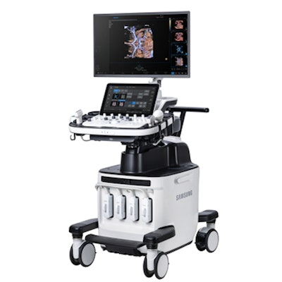
Samsung subsidiary Boston Imaging has added a new premier offering to its Hera line of ultrasound scanners for obstetrics and gynecology applications, the company said.
Boston Imaging's new Hera W10 Elite ultrasound scanner for ob/gyn applications. Photo courtesy of Samsung.Hera W10 Elite features a suite of artificial intelligence (AI) tools and clinical applications, according to the vendor. The new system features a number of updates compared with its predecessor: Hera W10. These include the following:
- Advanced MV-Flow: A tool that displays blood flow intensity by providing 2D and 3D visualization of microcirculatory and slow blood.
- Larger OLED display: The display area has been increased by 57%, while the OLED screen is designed to realistically represent black colors to make it well-suited for diverse ultrasound image characteristics, according to the vendor.
- A new transducer: The EV2-124 enables visualization of the fetal heart and brain, beginning at eight weeks endovaginally.
The company said it has also incorporated AI features such as HeartAssist for providing measurement results and automatically classifying ultrasound images into measurement views, ViewAssist for automatically classifying ultrasound images and annotating structures for measurement, and BiometryAssist for measuring fetal growth parameters with a single click while maintaining exam consistency.









![Examples of ultrasound findings and techniques. (A) Images in a 39-year-old male patient with a mass in the left thigh. The mass is heterogeneous on the B-mode US image (compared with the patient in D) and showed increased microvascularity (superb microvascular imaging [SMI]) and shear-wave elastography (SWE) values. Undifferentiated pleomorphic sarcoma was diagnosed at biopsy (with pleomorphic rhabdomyosarcoma in surgical specimen). (B) Images in an 18-year-old male patient with a mass in the left leg. The mass is hypoechoic on the B-mode image, with no other findings suggestive of malignancy. The lesion is in contact with the cortex of the tibia, which is slightly irregular. CT revealed a doubtful anteromedial tibial erosion. The microvascular study demonstrated high vascularization, suggestive of malignancy. Periosteal Ewing sarcoma was diagnosed with both histologic and immunohistochemical confirmation. (C) Images in a 69-year-old female patient with a lump growing on the outside of the left leg. Multiple SWE examinations were performed (please note the high values obtained in the measurements, whereas the color map highlights the stiffness relative to adjacent tissues). SMI showed areas of increased vascularization to target for sampling. Undifferentiated spindle cell sarcoma was diagnosed at biopsy, with residual leiomyosarcoma in the surgical specimen after neoadjuvant therapy. (D) Images in a 56-year-old female patient with a mass in the right thigh. The mass is heterogeneous at both B-mode ultrasound (similar to patient A) and MRI (coronal T2-weighted spectral attenuated inversion recovery [SPAIR]; T1-weighted pre-contrast and postcontrast imaging), which even shows uptake after the administration of paramagnetic contrast material, which is traditionally suggestive of malignancy. Low values at SMI and elastography are suggestive of benignity. Spindle cell lipoma was diagnosed at biopsy, with atypical spindle cell lipomatous tumor in the surgical specimen.](https://img.auntminnie.com/mindful/smg/workspaces/default/uploads/2026/08/images-radiol250278fig2.APCFLSvX6p.jpg?auto=format%2Ccompress&fit=crop&h=112&q=70&w=112)


