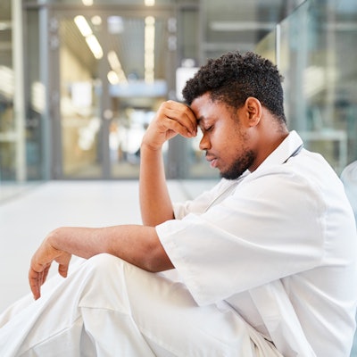
The phenomenon of burnout among radiology staff isn't new, but the COVID-19 pandemic has added an extra layer to it, two July 15 presentations delivered at ECR 2022 showed.
Dávid Sipos of the University of Pécs in Hungary detailed results from a survey he and colleagues conducted last year that showed the effects of the COVID-19 crisis on radiographer well-being, while Emily Skelton of City University of London outlined the pandemic's effect on obstetric sonographers.
Both presenters' findings corroborated recent research -- for example, a 2021 study published in Radiography concluded that the pandemic has caused anxiety and workplace-related stress, particularly among radiographers and a talk delivered in 2020 at AuntMinnie.com's Fall 2020 Virtual Conference (AMVC) highlighted the negative effects of coping with COVID-19.
Sipos' research consisted of survey data collected from 439 Society of Hungarian Radiographers members. The survey incorporated the Effort-Reward Imbalance and Maslach Burnout Inventory questionnaires along with further questions developed by Sipos and colleagues; it was distributed between January and March 2021. (The Effort-Reward Imbalance assesses how a mismatch between high effort expended and low rewards received at work affects employee health and well-being, while the Maslach inventory measures emotional exhaustion, depersonalization, and the sense of personal accomplishment).
His team found the following characteristics contributed to higher stress levels among radiographers during the COVID-19 pandemic:
- Being male
- Being younger (39 years old and below)
- Working in the healthcare system 20 years or less
- Being in quarantine during the pandemic
Those radiographers who interacted with confirmed or suspected COVID-19 patients and also worked in the emergency department reported higher levels of depersonalization and emotional exhaustion, Sipos noted. Being in quarantine significantly lowered a sense of personal achievement and raised emotional exhaustion, he said.
In her talk, Skelton described research that included data gathered from a survey distributed to 89 sonographers, the majority of whom were women (96%); almost half were working full-time for the National Health Service (49%). The survey was based on the Oldenburg Burnout Inventory, which consists of 16 statements that explore the state of respondents' "exhaustion-vigor and cynicism-dedication."
Skelton and colleagues found that 92% of survey respondents met burnout thresholds for exhaustion and 91% for "disengagement." Changes in role satisfaction correlated with survey respondents' perception of their own vulnerability to contracting COVID-19 in the course of their work with patients. Of the study participants, 65% stated that they were considering leaving the profession or changing their area of practice or working hours within five years.
How can this burnout among obstetric sonographers be addressed going forward? It's clear that proactive measures are needed, or workforce shortages caused by staff stress leaving their positions could impact patient care, Skelton said.
"Job-specific interventions are required to mitigate sonographer burnout and its consequences on workforce shortages and service provision beyond the COVID-19 pandemic," she concluded.