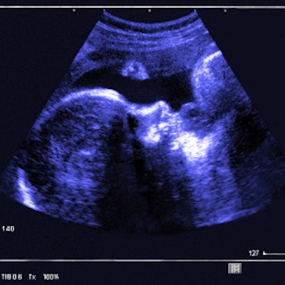
Congenital defects and lack of intrauterine heartbeat are leading factors in terminating pregnancies, and both can be detected on ultrasound scans, suggests a study published June 30 in the Taiwanese Journal of Obstetrics and Gynecology.
Researchers led by Dr. Ching Hua Hsiao from the National Yang Ming Chiao Tung University wrote that their findings can better inform obstetricians for planning and counseling regarding factors underlying decisions to induce intrauterine fetal death.
"It is important to educate all women about family planning to prevent large numbers of unwanted and unsafe pregnancy terminations," Hsiao and colleagues wrote.
Fetal death has several causes, including congenital chromosomal or structural abnormalities, elective termination of pregnancies, premature rupturing of fetal membranes, and cervical incompetence, as well as spontaneous missed abortion or accidental intrauterine fetal death.
A stillbirth occurs when the fetus dies at or after the 20th week of pregnancy. Determining the exact cause of stillbirths can be challenging. Researchers wrote that developing ways to improve determining causes and communication with patients and families can advance the understanding of stillbirth, as well as alleviate patient burden. Ultrasound can help identify structural abnormalities around the 20-week mark.
Termination of pregnancy is a controversial topic worldwide. The U.S. Supreme Court in June voted in favor of holding that the U.S. Constitution does not grant the right to abortion, overruling both Roe v. Wade and Planned Parenthood v. Casey. In Taiwan, meanwhile, girls younger than 20 years old need parental consent to terminate pregnancy.
Congenital defects can make fetal survival outside of the uterus impossible or have a poor prognosis, and babies born too prematurely may not survive before 23 weeks. Pregnancies are considered "nonviable" when the fetus no longer has a heartbeat. The Hsiao team wanted to identify the factors and frequencies of induced termination of pregnancies, using retrospective data. They looked at 488 terminations of pregnancies performed between January 2011 and December 2021.
All patients had been hospitalized to manage the induction of labor. Patient ages ranged from 12 to 46 years, with a median maternal age of 34. Out of the total, 52.2% of women had at least one prior delivery.
| Causes of fetal death | |
| Chromosomal and genetic abnormalities | 29.9% |
| No intrauterine heartbeats | 25.8% |
| Structural anomalies | 17.2% |
| Elective termination | 11.5% |
| Preterm premature rupture of membranes | 8.6% |
| Cervical incompetence | 6.6% |
| Other conditions | 0.4% |
Researchers also found that the gestational age in intrauterine fetal death was greater than other groups (p < 0.05), and the maternal age in the group of elective termination of pregnancies was younger than in other groups (p < 0.05).
They also found no significant changes from 2011 to 2021 for the rate of fetal deaths and stillbirths after excluding elective terminations.
The study authors wrote that in the 56 elective pregnancy termination cases (average age, 26.1 years), the three main reasons for termination were rape, too young to be a mother, and multiple births. The medial gestational ages for pregnancy termination due to congenital and structural abnormalities were 18.4 and 20.6 weeks, respectively. The authors added that it’s around this time that ultrasound scans are used to evaluate structural anomalies.
Hsiao and colleagues wrote that their findings support the need for improved methods to determine the cause of fetal death. "Development of novel postmortem examination to objectively determine the cause of fetal death is largely beneficial for both the clarification and prevention of intrauterine fetal death in the future," Hsiao and colleagues added.



![Examples of ultrasound findings and techniques. (A) Images in a 39-year-old male patient with a mass in the left thigh. The mass is heterogeneous on the B-mode US image (compared with the patient in D) and showed increased microvascularity (superb microvascular imaging [SMI]) and shear-wave elastography (SWE) values. Undifferentiated pleomorphic sarcoma was diagnosed at biopsy (with pleomorphic rhabdomyosarcoma in surgical specimen). (B) Images in an 18-year-old male patient with a mass in the left leg. The mass is hypoechoic on the B-mode image, with no other findings suggestive of malignancy. The lesion is in contact with the cortex of the tibia, which is slightly irregular. CT revealed a doubtful anteromedial tibial erosion. The microvascular study demonstrated high vascularization, suggestive of malignancy. Periosteal Ewing sarcoma was diagnosed with both histologic and immunohistochemical confirmation. (C) Images in a 69-year-old female patient with a lump growing on the outside of the left leg. Multiple SWE examinations were performed (please note the high values obtained in the measurements, whereas the color map highlights the stiffness relative to adjacent tissues). SMI showed areas of increased vascularization to target for sampling. Undifferentiated spindle cell sarcoma was diagnosed at biopsy, with residual leiomyosarcoma in the surgical specimen after neoadjuvant therapy. (D) Images in a 56-year-old female patient with a mass in the right thigh. The mass is heterogeneous at both B-mode ultrasound (similar to patient A) and MRI (coronal T2-weighted spectral attenuated inversion recovery [SPAIR]; T1-weighted pre-contrast and postcontrast imaging), which even shows uptake after the administration of paramagnetic contrast material, which is traditionally suggestive of malignancy. Low values at SMI and elastography are suggestive of benignity. Spindle cell lipoma was diagnosed at biopsy, with atypical spindle cell lipomatous tumor in the surgical specimen.](https://img.auntminnie.com/mindful/smg/workspaces/default/uploads/2026/08/images-radiol250278fig2.APCFLSvX6p.jpg?auto=format%2Ccompress&fit=crop&h=100&q=70&w=100)


![Examples of ultrasound findings and techniques. (A) Images in a 39-year-old male patient with a mass in the left thigh. The mass is heterogeneous on the B-mode US image (compared with the patient in D) and showed increased microvascularity (superb microvascular imaging [SMI]) and shear-wave elastography (SWE) values. Undifferentiated pleomorphic sarcoma was diagnosed at biopsy (with pleomorphic rhabdomyosarcoma in surgical specimen). (B) Images in an 18-year-old male patient with a mass in the left leg. The mass is hypoechoic on the B-mode image, with no other findings suggestive of malignancy. The lesion is in contact with the cortex of the tibia, which is slightly irregular. CT revealed a doubtful anteromedial tibial erosion. The microvascular study demonstrated high vascularization, suggestive of malignancy. Periosteal Ewing sarcoma was diagnosed with both histologic and immunohistochemical confirmation. (C) Images in a 69-year-old female patient with a lump growing on the outside of the left leg. Multiple SWE examinations were performed (please note the high values obtained in the measurements, whereas the color map highlights the stiffness relative to adjacent tissues). SMI showed areas of increased vascularization to target for sampling. Undifferentiated spindle cell sarcoma was diagnosed at biopsy, with residual leiomyosarcoma in the surgical specimen after neoadjuvant therapy. (D) Images in a 56-year-old female patient with a mass in the right thigh. The mass is heterogeneous at both B-mode ultrasound (similar to patient A) and MRI (coronal T2-weighted spectral attenuated inversion recovery [SPAIR]; T1-weighted pre-contrast and postcontrast imaging), which even shows uptake after the administration of paramagnetic contrast material, which is traditionally suggestive of malignancy. Low values at SMI and elastography are suggestive of benignity. Spindle cell lipoma was diagnosed at biopsy, with atypical spindle cell lipomatous tumor in the surgical specimen.](https://img.auntminnie.com/mindful/smg/workspaces/default/uploads/2026/08/images-radiol250278fig2.APCFLSvX6p.jpg?auto=format%2Ccompress&fit=crop&h=112&q=70&w=112)