
UltraSight highlighted a study presented at the annual conference of the Israel Heart Society in Tel Aviv that showed its artificial intelligence (AI) software for guiding real-time ultrasound imaging can be employed by novice users.
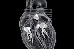
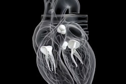
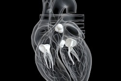
UltraSight's software pairs with point-of-care ultrasound devices to analyze ultrasound video streams and provides users with real-time guidance on how to maneuver the ultrasound probe to obtain diagnostic quality cardiac images, such as 2D transthoracic echocardiography images.
In the study, three novice medical assistants, none of whom had any cardiac ultrasound experience, underwent an eight-hour training course before scanning 61 cardiac patients in all 10 standard echocardiography views. The medical assistants' exams successfully permitted the assessment of left ventricular size and function, right ventricular size, and presence of significant pericardial effusion in 100% of patients, the company said.
UltraSight's novel technology may enable novice users, after minimal training, to perform high-quality diagnostic echocardiographic studies, concluded Dr. Robert Klempfner, director of the Cardiac Rehabilitation Institute at Sheba Medical Center in Tel Aviv, who led the study.
Ultimately, the technology has the potential to increase user adoption of point-of-care ultrasound and accelerate patient care in a variety of clinical settings, UltraSight said.







![Examples of ultrasound findings and techniques. (A) Images in a 39-year-old male patient with a mass in the left thigh. The mass is heterogeneous on the B-mode US image (compared with the patient in D) and showed increased microvascularity (superb microvascular imaging [SMI]) and shear-wave elastography (SWE) values. Undifferentiated pleomorphic sarcoma was diagnosed at biopsy (with pleomorphic rhabdomyosarcoma in surgical specimen). (B) Images in an 18-year-old male patient with a mass in the left leg. The mass is hypoechoic on the B-mode image, with no other findings suggestive of malignancy. The lesion is in contact with the cortex of the tibia, which is slightly irregular. CT revealed a doubtful anteromedial tibial erosion. The microvascular study demonstrated high vascularization, suggestive of malignancy. Periosteal Ewing sarcoma was diagnosed with both histologic and immunohistochemical confirmation. (C) Images in a 69-year-old female patient with a lump growing on the outside of the left leg. Multiple SWE examinations were performed (please note the high values obtained in the measurements, whereas the color map highlights the stiffness relative to adjacent tissues). SMI showed areas of increased vascularization to target for sampling. Undifferentiated spindle cell sarcoma was diagnosed at biopsy, with residual leiomyosarcoma in the surgical specimen after neoadjuvant therapy. (D) Images in a 56-year-old female patient with a mass in the right thigh. The mass is heterogeneous at both B-mode ultrasound (similar to patient A) and MRI (coronal T2-weighted spectral attenuated inversion recovery [SPAIR]; T1-weighted pre-contrast and postcontrast imaging), which even shows uptake after the administration of paramagnetic contrast material, which is traditionally suggestive of malignancy. Low values at SMI and elastography are suggestive of benignity. Spindle cell lipoma was diagnosed at biopsy, with atypical spindle cell lipomatous tumor in the surgical specimen.](https://img.auntminnie.com/mindful/smg/workspaces/default/uploads/2026/08/images-radiol250278fig2.APCFLSvX6p.jpg?auto=format%2Ccompress&fit=crop&h=112&q=70&w=112)