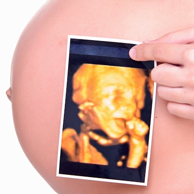
Researchers have developed a new ultrasound technique to monitor the placenta for impaired fetal blood flow early in pregnancy, according to study funded by the U.S. National Institutes of Health and published in April in eBioMedicine.
The study, led by John G. Sled, PhD, from the Hospital for Sick Children in Toronto, found that by using conventional ultrasound equipment, subtle differences in the pulsation of fetal blood can be found through the arteries at the fetal and placental ends of the umbilical cord. This potentially enables physicians to identify placental abnormalities that impair fetal blood flow and, if necessary, deliver the fetus early.
Like current ultrasound techniques, the new technique can also detect impaired flow of maternal blood through the placenta, authors said.
Researchers tested the new technique with ultrasound scans on women between the 26th and 32nd weeks of pregnancy and diagnosed those with circulatory problems in the maternal or fetal part of the placenta.
The authors say their method provides a way to diagnose circulation problems in the placenta that can harm the fetus and would otherwise go undetected until late in pregnancy.







![Examples of ultrasound findings and techniques. (A) Images in a 39-year-old male patient with a mass in the left thigh. The mass is heterogeneous on the B-mode US image (compared with the patient in D) and showed increased microvascularity (superb microvascular imaging [SMI]) and shear-wave elastography (SWE) values. Undifferentiated pleomorphic sarcoma was diagnosed at biopsy (with pleomorphic rhabdomyosarcoma in surgical specimen). (B) Images in an 18-year-old male patient with a mass in the left leg. The mass is hypoechoic on the B-mode image, with no other findings suggestive of malignancy. The lesion is in contact with the cortex of the tibia, which is slightly irregular. CT revealed a doubtful anteromedial tibial erosion. The microvascular study demonstrated high vascularization, suggestive of malignancy. Periosteal Ewing sarcoma was diagnosed with both histologic and immunohistochemical confirmation. (C) Images in a 69-year-old female patient with a lump growing on the outside of the left leg. Multiple SWE examinations were performed (please note the high values obtained in the measurements, whereas the color map highlights the stiffness relative to adjacent tissues). SMI showed areas of increased vascularization to target for sampling. Undifferentiated spindle cell sarcoma was diagnosed at biopsy, with residual leiomyosarcoma in the surgical specimen after neoadjuvant therapy. (D) Images in a 56-year-old female patient with a mass in the right thigh. The mass is heterogeneous at both B-mode ultrasound (similar to patient A) and MRI (coronal T2-weighted spectral attenuated inversion recovery [SPAIR]; T1-weighted pre-contrast and postcontrast imaging), which even shows uptake after the administration of paramagnetic contrast material, which is traditionally suggestive of malignancy. Low values at SMI and elastography are suggestive of benignity. Spindle cell lipoma was diagnosed at biopsy, with atypical spindle cell lipomatous tumor in the surgical specimen.](https://img.auntminnie.com/mindful/smg/workspaces/default/uploads/2026/08/images-radiol250278fig2.APCFLSvX6p.jpg?auto=format%2Ccompress&fit=crop&h=112&q=70&w=112)


