Ultrasound-guided tunneled central venous access proved safe for children in a randomized clinical trial published on December 24 in the Journal of Surgical Research. The trial included more than 100 patients and showed that ultrasound-guided placement reduced operating time and preserved vein size.
Ultrasound-guided line placement is already the standard of practice in adults, and it's becoming an increasingly popular option for children. The authors believed the new trial was the first to directly compare ultrasound-guided line placement with open surgery, and their findings help put to rest any concerns over the use of the technique in a pediatric population.
"This study demonstrated ultrasound-guided percutaneous insertion was safe, resulted in shorter operating times, no increased risk of complication rates, and preservation of vessel size," wrote the authors, led by Dr. Soundappan Soundappan, a general pediatric surgeon and head of trauma at the Children's Hospital at Westmead in Sydney, Australia.
The study included 108 patients, ranging in age from 9 months to 17 years, who had been referred to the surgery department for insertion of a cuffed double-lumen central line or an implantable vascular access device (IVAD). A computer program randomly assigned the patients to undergo placement through open surgery or an ultrasound-guided procedure.
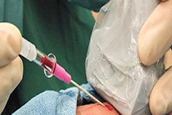
For patients randomized to the ultrasound group, surgeons with varying levels of experience performed a percutaneous technique using the Sonosite M-Turbo system from Fujifilm Sonosite with an intraoperative linear array probe. Under ultrasound guidance, they passed the needle perpendicular to the transducer axis and in line with the internal jugular vein (IJV), then performed the vein puncture.
| Ultrasound guidance vs. open surgery for central venous access in children | |||
| Open surgery | Ultrasound-guided procedure | p-value | |
| Time | 44 minutes | 56 minutes | 0.008 |
| Vein size after removal | 9.96 mm | 6.64 mm | 0.002 |
| Percentage of children with immediate complications | 8% | 14% | 0.356 |
| Percentage of children with late complications | 10% | 13% | 0.610 |
Ultrasound-guided placement took significantly less time than open surgery. In addition, patients in the ultrasound group had significantly larger vein size after removal, despite having similar vein sizes before line or IVAD insertion.
Furthermore, patients in both groups experienced similar complication rates. In both groups, the need for more than one insertion was the most common immediate complication, which occurred for five patients in the ultrasound group and seven in the open surgery group.
A similar proportion of patients in both groups also experienced late complications. The most common complication was infection, which occurred in seven children in the ultrasound group and six children in the open surgery group. One child in the ultrasound group also experienced a blocked catheter.
While the results are promising, the authors said the ultrasound-guided procedure didn't save as much time as they had hoped for, likely because a radiology professional was not part of the research team. Radiologists and technologists were also often committed to another procedure, leading to delays in the operating room.
"Position of the line was confirmed by on-table fluoroscopy but there was no dedicated radiology technician present in the operating room at the time of the procedure," the author wrote. They later added, "Absence of a dedicated radiology technician to the lines list meant our times were longer than they should be."
However, even with a shortage of radiology team members, the ultrasound-guided placement provided its worth in the trial, and the surgery department has come to rely on ultrasound-guidance in their day-to-day practice. In fact, the hospital now uses the ultrasound-guided percutaneous technique as the preferred choice for children who need a central line or IVAD placement.
"The study as it was performed was reflective of real-life practice and showed ultrasound-guided percutaneous lines were safe to perform," the authors concluded.