Researchers from Japan have developed a blood pressure monitor that utilizes a tiny ultrasound probe instead of compression to monitor systolic and diastolic changes. The science behind the prototype was presented as a virtual poster at the 2020 European Society of Cardiology (ESC) Conference.
Lead author Dr. Kazunori Uemura, PhD, and his team from the National Cerebral and Cardiovascular Center in Japan designed the ultrasound-based monitor to unobtrusively track blood pressure on-the-go. Once calibrated, the ultrasound-based device produced blood pressure readings comparable to conventional technology in dogs.
"This method reliably tracks [blood pressure] changes without occlusive cuff inflation," the authors wrote in their poster abstract.
Monitoring blood pressure out-of-the-office and at regular intervals is important for hypertension diagnosis and management, but current compression-based blood pressure monitors can interfere with the daily lives of patients. Uemura and colleagues saw ultrasound as a potential alternative to compression-based technology.
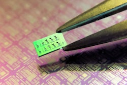
The cuff they created utilizes a tiny ultrasound probe about half the size of a box of matches. The probe sits snugly in between a patient's skin and the larger blood pressure cuff.
Upon mild cuff inflation, the ultrasound transducer's 65 piezoelectric elements measure changes to an artery situated underneath the probe. Uemura and colleagues use these measurements to calculate estimated diastolic and systolic blood pressure.
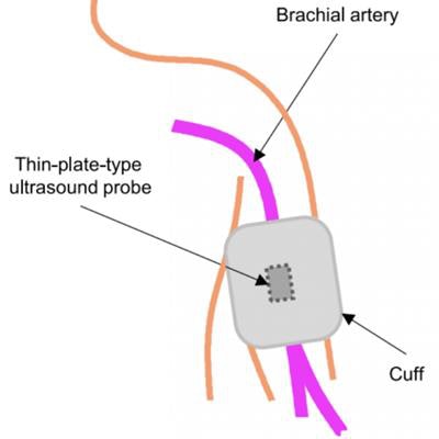
 (A) Schematic illustration of the cuff and ultrasound probe wrapped around the upper arm and positioned over the brachial artery. (B) Longitudinal cross section illustration of the ultrasound probe and inflated cuff. When the cuff is inflated, the device tracks the ultrasonic dimension of the brachial artery. Image courtesy of Dr. Kazunori Uemura, PhD.
The authors tested the cuff's accuracy for blood pressure monitoring on the right femoral artery of six anesthetized dogs. Measurements taken with the ultrasound-based device correlated strongly with compression-based readings, according to coefficient of determination (R2) analysis.
With just one-time calibration, the diastolic blood pressure readings had a bias of 3.9 ± 7.9 mmHg. This measurement fell within the acceptable bias range of < 5 ± < 8 mmHg set by the Association for the Advancement of Medical Instrumentation (AAMI).
However, with one-time calibration, the systolic blood pressure readings had a bias of 2.6 ± 18.9 mmHg, outside of the AAMI acceptable range. To improve the systolic readings, the authors added machine learning and a support vector algorithm to the initial, one-time calibration. The new analysis yielded systolic measurements with a bias of 0.7 ± 6.9 mmHg, meeting the AAMI guidelines.
"Once calibrated, this method measures [diastolic blood pressure] accurately," the authors wrote. "With the aid of machine learning, precision in [systolic blood pressure] prediction was greatly improved to acceptable levels."
This type of ultrasound-based technology is a long way from use in the clinic, and the research team still has yet to test the device's accuracy on people. But if the cuff holds up in further testing, the combination of ultrasound and machine learning could lead to better mobile blood pressure monitoring.
"This method with machine learning approach has potential for stress-free [blood pressure] measurement in ambulatory [blood pressure] monitoring," the authors concluded.