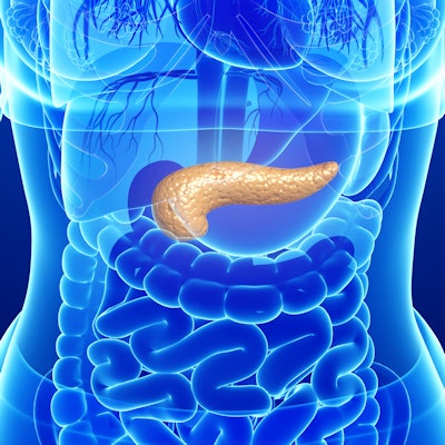
A new "virtual biopsy" using ultrasound guidance allows doctors to definitively diagnose cysts in the pancreas with 97% accuracy, enabling them to remove precancerous cysts early, according to researchers from Ohio State University.
Many patients are diagnosed with pancreatic cysts incidentally when undergoing an MRI or CT scan for another indication, and patients are usually asymptomatic until the cancer is advanced, making treatment challenging, the researchers noted.
The current standard for biopsies involves testing the fluid inside cysts to determine whether they're cancerous with 71% accuracy. When using virtual biopsy, diagnostic accuracy jumps to 97%.
Virtual biopsy uses endoscopic ultrasound-guided needle-based confocal laser endomicroscopy. The research team from Ohio State University in Columbus led by Dr. Somashekar Krishna performed a prospective study of 144 consecutive patients with a suspected pancreatic cystic lesion. They found endoscopic ultrasound-guided needle-based confocal laser endomicroscopy identified mucinous cystic lesions with 98% sensitivity, 94% specificity, and 97% accuracy. They presented their findings at the recent American Pancreatic Association's annual meeting.
The Ohio State researchers are now working to train doctors nationwide to perform this new diagnostic method and read the images provided by the scope. They're also developing artificial intelligence that will flag cases that are likely precancerous.









![Examples of ultrasound findings and techniques. (A) Images in a 39-year-old male patient with a mass in the left thigh. The mass is heterogeneous on the B-mode US image (compared with the patient in D) and showed increased microvascularity (superb microvascular imaging [SMI]) and shear-wave elastography (SWE) values. Undifferentiated pleomorphic sarcoma was diagnosed at biopsy (with pleomorphic rhabdomyosarcoma in surgical specimen). (B) Images in an 18-year-old male patient with a mass in the left leg. The mass is hypoechoic on the B-mode image, with no other findings suggestive of malignancy. The lesion is in contact with the cortex of the tibia, which is slightly irregular. CT revealed a doubtful anteromedial tibial erosion. The microvascular study demonstrated high vascularization, suggestive of malignancy. Periosteal Ewing sarcoma was diagnosed with both histologic and immunohistochemical confirmation. (C) Images in a 69-year-old female patient with a lump growing on the outside of the left leg. Multiple SWE examinations were performed (please note the high values obtained in the measurements, whereas the color map highlights the stiffness relative to adjacent tissues). SMI showed areas of increased vascularization to target for sampling. Undifferentiated spindle cell sarcoma was diagnosed at biopsy, with residual leiomyosarcoma in the surgical specimen after neoadjuvant therapy. (D) Images in a 56-year-old female patient with a mass in the right thigh. The mass is heterogeneous at both B-mode ultrasound (similar to patient A) and MRI (coronal T2-weighted spectral attenuated inversion recovery [SPAIR]; T1-weighted pre-contrast and postcontrast imaging), which even shows uptake after the administration of paramagnetic contrast material, which is traditionally suggestive of malignancy. Low values at SMI and elastography are suggestive of benignity. Spindle cell lipoma was diagnosed at biopsy, with atypical spindle cell lipomatous tumor in the surgical specimen.](https://img.auntminnie.com/mindful/smg/workspaces/default/uploads/2026/08/images-radiol250278fig2.APCFLSvX6p.jpg?auto=format%2Ccompress&fit=crop&h=112&q=70&w=112)


