Augmented reality (AR) may improve the performance of ultrasound image guidance, say researchers from Canada who developed a new technique using AR and ultrasound to help pilot needle insertion into the spine. They discuss their method in an article recently published in Ultrasound in Medicine and Biology.
The group developed a novel method using a combination of AR and ultrasound to guide epidural injections, the most common intervention in pain management of the spine (Ultrasound Med Biol, July 4, 2019).
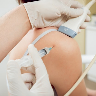
Clinicians typically perform epidural injections by relying on anatomic landmarks to guide needle insertion, a blind technique that suffers from a low first-pass success rate, noted first author Golafsoun Ameri, PhD, from the Robarts Research Institute, and colleagues. Recently, various groups have attempted to use imaging modalities such as ultrasound to improve needle navigation and placement, but the visualization provided by 2D imaging is limited in depth and clarity.
"Despite promising results, real-time ultrasound guidance for needle navigation and placement in the epidural space remains challenging and has not been adopted for everyday practice ... largely because of poor needle visibility ... and poor target visibility," they wrote.
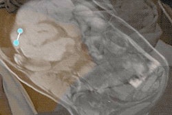
To address these challenges, Ameri and colleagues integrated AR technology into ultrasound image guidance using proprietary software that they developed on two open-source software platforms (3D Slicer; Public Library for UltraSound). The software enabled them to overlay virtual 3D models, or AR models, onto a display showing ultrasound images in real-time.
After calibrating the ultrasound probe, the researchers had four clinicians test the viability of their combined AR and ultrasound image guidance technique in a series of trials. For the trials, the clinicians used either AR with ultrasound or ultrasound alone to guide needle insertion into a 3D-printed spine model that they had previously created based on patient CT scans.
The researchers found that all seven of the AR-guided procedures were successful, whereas nearly half of the attempts using only ultrasound guidance resulted in accidental punctures of the dura mater of the spinal cord. What's more, the AR-guided method took a shorter amount of time, required less effort, and was associated with a lower level of subjective "frustration" than ultrasound guidance, according to the clinicians.
| Effectiveness of AR + ultrasound vs. ultrasound alone for spine needle insertion | ||
| Ultrasound guidance | AR + ultrasound guidance | |
| Rate for accurate needle insertion | 57% | 100% |
| Procedure time | ~58 seconds | ~34 seconds |
Though only a small-scale evaluation, the study's findings demonstrate the potential of AR plus ultrasound image guidance to improve needle placement on several fronts -- from making the task seem more intuitive to reducing procedural time and intraoperative complications, the authors noted.
"The AR ultrasound image guidance system was implemented on an open-source platform with the aim of fostering research collaboration and accelerating the improvement of state-of-the-art guidance systems for overcoming challenges in not only epidural injections but also other common needle interventions to achieve improved patient outcomes," Ameri and colleagues wrote.
In addition, the AR technique includes recording and playback functionalities that allow for the technique's use in skill assessment and training, they wrote. These features may promote the technology's eventual adoption in day-to-day clinical practice for a wide variety of clinical procedures, such as biopsy, brachytherapy, and minimally invasive cardiac procedures.