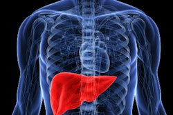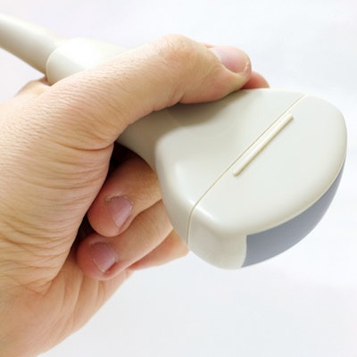
Two-dimensional shear-wave elastography (2D SWE) is an effective tool for identifying cirrhosis, according to a study by researchers from China and published online July 24 in Radiology.
The findings are good news for patients, since the technique could offer a less invasive alternative than the current practice of liver biopsy, wrote a team led by Dr. Yongyan Gao of the Chinese People's Liberation Army General Hospital in Beijing.
"Although liver biopsy remains the reference standard to assess liver fibrosis, it is limited by its invasiveness and potential complications," the group wrote. "Two-dimensional shear-wave elastography by using the supersonic shear imaging technique is the first ultrasound-based real-time 2D SWE imaging technique with several advantages."
Hepatitis B is a major public health problem, and reliable ways of evaluating the stages of liver fibrosis are necessary for treating patients with the disease, Gao and colleagues wrote. Because of liver biopsy's limitations, techniques such as transient elastography, which uses both ultrasound and low-frequency elastic waves to evaluate elasticity, and 2D shear-wave elastography have become increasingly attractive.
The researchers sought to evaluate 2D SWE's performance in predicting cirrhosis, comparing its results to those of serum marker data (the aspartate aminotransferase-to-platelet ratio index [APRI], which predicts hepatitis virus progression), the Fibrosis-4 index for estimating liver scarring, and transient elastography.
The study included 402 patients with chronic hepatitis B who underwent liver biopsy and 2D shear-wave elastography exams between January 2015 and January 2016. Of the total patient cohort, 154 had chronic infection and 248 had chronic hepatitis. The investigators used the area under the receiver operating characteristic curve (AUC) to measure performance.
Gao and colleagues found that the overall AUCs of 2D SWE for diagnosing significant fibrosis and cirrhosis were higher than those of the other techniques, and the modality was most effective in ruling in or out the presence of cirrhosis in patients with chronic infection -- which could have implications for earlier and more effective treatment, according to the team.
| Diagnostic accuracy of techniques for predicting cirrhosis | ||||
| Serum markers (APRI) | Liver scarring (Fibrosis-4) | Transient elastography | 2D SWE | |
| AUC | 0.70 | 0.73 | 0.80 | 0.87 |
| Sensitivity | 27.6% | 38.8% | 28.6% | 56.6% |
| Sensitivity | 89.7% | 90% | 89.5% | 90.1% |
"The performance of 2D SWE is superior to other noninvasive methods" and is effective for ruling in or out cirrhosis, concluded the group, adding that it "may be useful for the guidance of antiviral treatment."