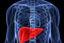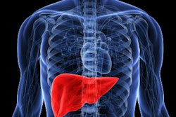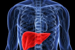
Transient elastography is an effective diagnostic tool for identifying liver fibrosis in children, according to a study from South Korea that was published in the November issue of the American Journal of Roentgenology.
Although transient elastography has been shown to be effective in adults, it has been unclear whether it would also be useful for children, wrote a team led by Dr. Jae-Yeon Hwang of Pusan National University Yangsan Hospital. Children develop chronic liver disease due to hepatitis, fatty liver disease, genetic disorders, or liver dysfunction caused by congestive heart failure. Transient elastography -- which uses both ultrasound and low-frequency elastic waves to evaluate tissue elasticity -- offers a noninvasive diagnostic alternative to liver biopsy.
"Accurate assessment and monitoring of liver fibrosis is important for making therapeutic plans and determining prognosis," Hwang and colleagues wrote. "Currently, the reference standard for assessing the severity of liver fibrosis is liver biopsy."
However, "ultrasound-based elastography has become a promising imaging modality for the measurement of liver stiffness in children because of its ease of access, noninvasiveness, and high accuracy," they noted.
Using Medline and Embase, the researchers identified 11 studies involving 723 patients that assessed the performance of transient elastography for diagnosing the severity of liver fibrosis in children and adolescents. The mean age of patients in these studies ranged from 2.5 months to 13 years (AJR, November 2018, Vol. 211:5, pp. W257-W266).
Hwang's team found that transient elastography had a sensitivity of 95% and a specificity of 90% for this particular application.
"This meta-analysis revealed that the summary sensitivity and specificity of transient elastography for the diagnosis of significant liver fibrosis in children ... were even higher than those of adult studies," the group noted. "This may be because children have thinner abdominal walls, which permits better transmission of shear waves into the liver."
But despite the positive study findings -- and the group's conclusion that transient elastography is an effective way to appraise liver fibrosis in pediatric patients -- it's crucial that radiologists keep a few limitations specific to this population in mind.
"In a real clinical practice, a number of factors may differ between adults and children, such as less patient cooperation, which might result in lower compliance, including motion and breathing status, and the smaller and thinner body size of children compared to adults," Hwang and colleagues noted.


![Examples of ultrasound findings and techniques. (A) Images in a 39-year-old male patient with a mass in the left thigh. The mass is heterogeneous on the B-mode US image (compared with the patient in D) and showed increased microvascularity (superb microvascular imaging [SMI]) and shear-wave elastography (SWE) values. Undifferentiated pleomorphic sarcoma was diagnosed at biopsy (with pleomorphic rhabdomyosarcoma in surgical specimen). (B) Images in an 18-year-old male patient with a mass in the left leg. The mass is hypoechoic on the B-mode image, with no other findings suggestive of malignancy. The lesion is in contact with the cortex of the tibia, which is slightly irregular. CT revealed a doubtful anteromedial tibial erosion. The microvascular study demonstrated high vascularization, suggestive of malignancy. Periosteal Ewing sarcoma was diagnosed with both histologic and immunohistochemical confirmation. (C) Images in a 69-year-old female patient with a lump growing on the outside of the left leg. Multiple SWE examinations were performed (please note the high values obtained in the measurements, whereas the color map highlights the stiffness relative to adjacent tissues). SMI showed areas of increased vascularization to target for sampling. Undifferentiated spindle cell sarcoma was diagnosed at biopsy, with residual leiomyosarcoma in the surgical specimen after neoadjuvant therapy. (D) Images in a 56-year-old female patient with a mass in the right thigh. The mass is heterogeneous at both B-mode ultrasound (similar to patient A) and MRI (coronal T2-weighted spectral attenuated inversion recovery [SPAIR]; T1-weighted pre-contrast and postcontrast imaging), which even shows uptake after the administration of paramagnetic contrast material, which is traditionally suggestive of malignancy. Low values at SMI and elastography are suggestive of benignity. Spindle cell lipoma was diagnosed at biopsy, with atypical spindle cell lipomatous tumor in the surgical specimen.](https://img.auntminnie.com/mindful/smg/workspaces/default/uploads/2026/08/images-radiol250278fig2.APCFLSvX6p.jpg?auto=format%2Ccompress&fit=crop&h=100&q=70&w=100)


![Examples of ultrasound findings and techniques. (A) Images in a 39-year-old male patient with a mass in the left thigh. The mass is heterogeneous on the B-mode US image (compared with the patient in D) and showed increased microvascularity (superb microvascular imaging [SMI]) and shear-wave elastography (SWE) values. Undifferentiated pleomorphic sarcoma was diagnosed at biopsy (with pleomorphic rhabdomyosarcoma in surgical specimen). (B) Images in an 18-year-old male patient with a mass in the left leg. The mass is hypoechoic on the B-mode image, with no other findings suggestive of malignancy. The lesion is in contact with the cortex of the tibia, which is slightly irregular. CT revealed a doubtful anteromedial tibial erosion. The microvascular study demonstrated high vascularization, suggestive of malignancy. Periosteal Ewing sarcoma was diagnosed with both histologic and immunohistochemical confirmation. (C) Images in a 69-year-old female patient with a lump growing on the outside of the left leg. Multiple SWE examinations were performed (please note the high values obtained in the measurements, whereas the color map highlights the stiffness relative to adjacent tissues). SMI showed areas of increased vascularization to target for sampling. Undifferentiated spindle cell sarcoma was diagnosed at biopsy, with residual leiomyosarcoma in the surgical specimen after neoadjuvant therapy. (D) Images in a 56-year-old female patient with a mass in the right thigh. The mass is heterogeneous at both B-mode ultrasound (similar to patient A) and MRI (coronal T2-weighted spectral attenuated inversion recovery [SPAIR]; T1-weighted pre-contrast and postcontrast imaging), which even shows uptake after the administration of paramagnetic contrast material, which is traditionally suggestive of malignancy. Low values at SMI and elastography are suggestive of benignity. Spindle cell lipoma was diagnosed at biopsy, with atypical spindle cell lipomatous tumor in the surgical specimen.](https://img.auntminnie.com/mindful/smg/workspaces/default/uploads/2026/08/images-radiol250278fig2.APCFLSvX6p.jpg?auto=format%2Ccompress&fit=crop&h=112&q=70&w=112)